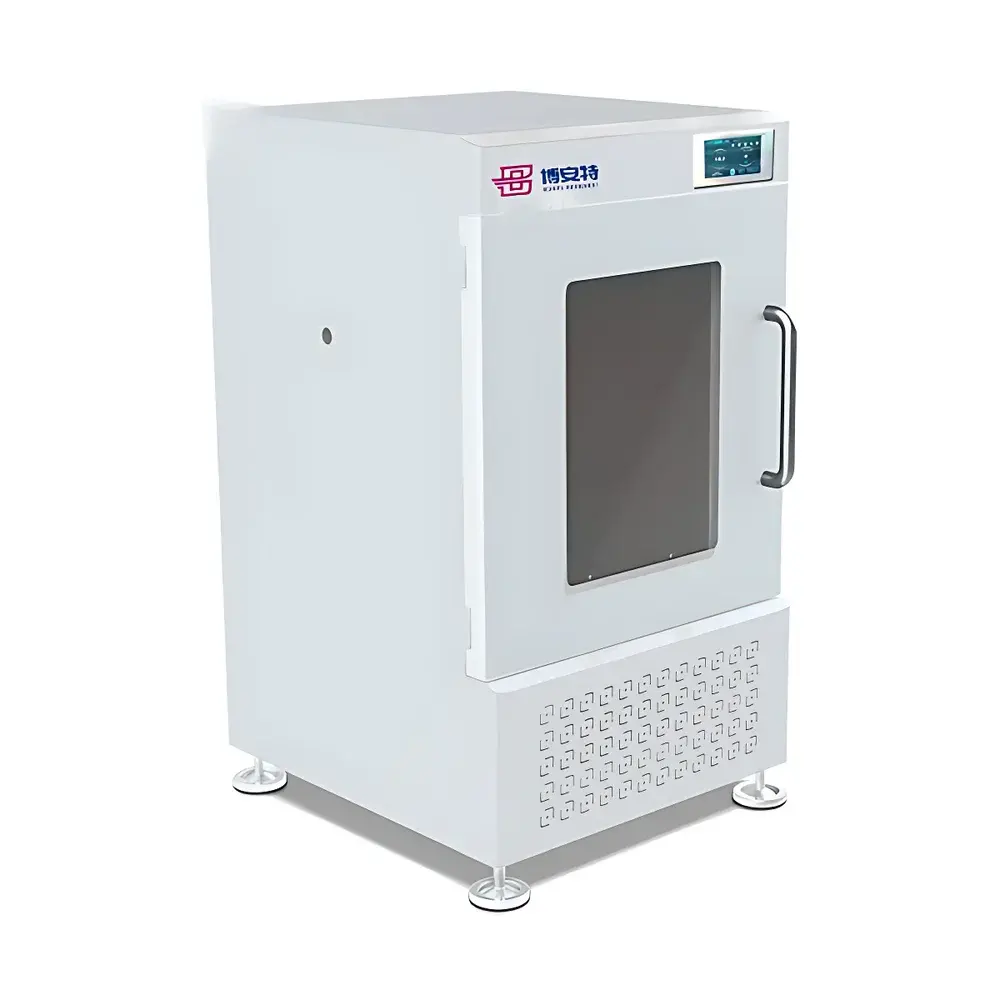
IVIM Two-Photon Microscope IVM-MS
| Origin | South Korea |
|---|---|
| Manufacturer Type | Authorized Distributor |
| Origin Category | Imported |
| Model | IVM-MS |
| Price Range | USD 420,000 – 700,000 |
| Instrument Type | Optical Imaging System |
| Energy Resolution | 0.2 keV |
| Scan Resolution | 1024 × 1024 pixels |
| Frame Rate | 100 fps |
| Field of View (FOV) | 100 × 100 µm² to 10 × 10 mm² |
| Sample Capacity | 1 animal per imaging session |
Overview
The IVIM Two-Photon Microscope IVM-MS is a high-performance, integrated in vivo optical imaging platform engineered for longitudinal, high-fidelity cellular and subcellular observation in awake or anesthetized small mammals. Based on the principle of near-infrared (NIR) two-photon excitation fluorescence (TPEF), the system leverages ultrafast femtosecond laser illumination (690–1050 nm tunable range) to achieve deep-tissue penetration up to 1000 µm with minimized phototoxicity and out-of-focus photobleaching. Unlike conventional confocal systems, the IVM-MS utilizes simultaneous dual-modal capability: a dedicated two-photon imaging path for structural and functional deep-tissue visualization, and an optional multi-channel confocal module supporting up to four user-selectable excitation wavelengths (including 405, 488, 561, and 642 nm) for complementary spectral flexibility. Its core architecture is optimized for quantitative intravital microscopy—enabling dynamic monitoring of physiological processes across spatial scales from single-cell motility to organ-level perfusion and immune trafficking.
Key Features
- Ultrafast resonant galvanometric scanning with rotating polygon mirror technology, enabling acquisition at 100 fps at 512 × 512 resolution and maintaining uniform excitation intensity across the full FOV—eliminating central signal attenuation and peripheral photobleaching artifacts.
- Integrated real-time motion artifact compensation powered by GPU-accelerated parallel computation; supports adaptive correction for both slow (e.g., hepatic, renal parenchyma) and rapid (e.g., cardiac, pulmonary) tissue dynamics without user intervention.
- Expandable modular design accommodating custom optical paths, including optional second-harmonic generation (SHG), third-harmonic generation (THG), and fluorescence lifetime imaging (FLIM) configurations.
- Motorized XYZ stage with sub-micron repeatability and piezo-driven objective positioning for precise z-stack acquisition and time-lapse volumetric reconstruction.
- Thermally stabilized environmental chamber (37°C ± 0.3°C) with CO2 and O2 regulation, compatible with chronic cranial window, abdominal imaging window, and dorsal skin-fold chamber preparations.
Sample Compatibility & Compliance
The IVM-MS is validated for use with murine models under standard vivarium conditions, supporting head-fixed, treadmill-based, or freely moving behavioral paradigms via lightweight head-mounted coupling optics. It complies with ISO 13485:2016 requirements for medical device-related research instrumentation and meets essential safety standards per IEC 60825-1 (laser product classification Class IV). All imaging protocols adhere to ARRIVE 2.0 guidelines and support GLP-compliant data traceability when paired with audit-trail-enabled software modules. The system is routinely deployed in facilities operating under AAALAC-accredited animal care programs and aligns with NIH Office of Laboratory Animal Welfare (OLAW) standards for in vivo imaging ethics and reporting.
Software & Data Management
Control and analysis are performed using IVIM-Studio v4.x—a native 64-bit application built on Qt and CUDA frameworks. The software provides synchronized hardware control, real-time motion-corrected preview, batch processing pipelines for drift correction, registration, and intensity normalization. Raw data are stored in standardized HDF5 format with embedded metadata (acquisition timestamp, laser power, PMT gain, objective magnification, and animal ID). Export options include TIFF stacks, NIfTI volumes, and CSV-formatted kinetic parameter tables. For regulatory environments, optional FDA 21 CFR Part 11 compliance packages are available, featuring electronic signatures, role-based access control, and immutable audit logs.
Applications
- Dynamic immunology: Tracking T-cell migration, dendritic cell–T-cell synapse formation, and neutrophil extracellular trap (NET) deployment in lymph nodes, bone marrow, and inflamed tissues.
- Neurovascular coupling studies: Simultaneous calcium imaging of neuronal ensembles and hemodynamic response mapping in cortex, hippocampus, and retina.
- Oncology research: Longitudinal monitoring of tumor cell intravasation/extravasation, angiogenic sprouting, and stromal remodeling in orthotopic and metastatic xenograft models (e.g., glioblastoma, melanoma, pancreatic ductal adenocarcinoma).
- Metabolic and fibrotic disease modeling: Quantitative assessment of hepatocyte lipid droplet dynamics, pancreatic β-cell mass turnover, and collagen fiber reorganization in liver/kidney fibrosis models.
- Developmental biology: Time-lapse imaging of embryonic organogenesis and postnatal vascular maturation using transgenic fluorescent reporters (e.g., Tie2-GFP, Cx3cr1-RFP).
FAQ
What is the maximum achievable imaging depth in scattering tissue?
Typical penetration depth reaches 800–1000 µm in murine cortical tissue or liver parenchyma using 920 nm excitation and high-numerical-aperture water-immersion objectives (25×, NA 1.05). Depth is modulated by tissue optical properties and laser pulse energy.
Can the system perform simultaneous two-photon and confocal imaging?
Yes—the IVM-MS features a dichroic beam combiner that enables concurrent acquisition from both modalities, allowing spectral unmixing of overlapping fluorophores (e.g., GFP + tdTomato + Cy5) with temporal synchronization within ±50 µs.
Is chronic imaging over multiple days supported?
The platform supports repeated imaging sessions over ≥14 days using stereotaxically anchored cranial windows or implantable abdominal imaging chambers, with automated focus stabilization and drift correction active during each session.
Does the system integrate with electrophysiology or optogenetics hardware?
Yes—standard TTL and analog I/O ports enable bidirectional synchronization with patch-clamp amplifiers, LED/laser optogenetic stimulators, and behavioral monitoring systems (e.g., rotary encoders, lickometers).
What training and service support is provided?
IVIM offers on-site installation, operator certification (3-day intensive course), and annual preventive maintenance contracts including laser recalibration, optical alignment verification, and software update deployment. Remote diagnostics and priority escalation are included in all enterprise support tiers.
Related Products