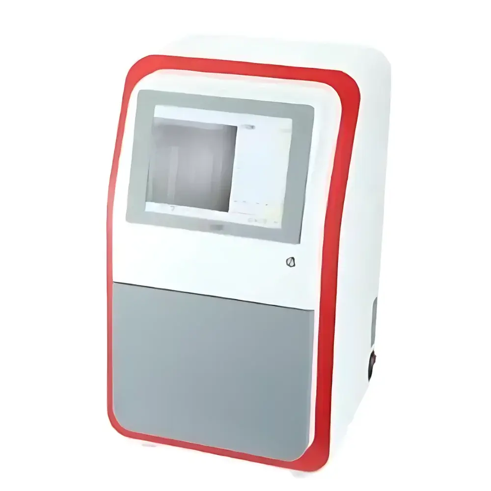
Jiapeng JP-K3600Plus Chemiluminescence Gel Imaging System
| Brand | Jiapeng |
|---|---|
| Origin | Shanghai, China |
| Manufacturer Type | Authorized Distributor |
| Product Category | Domestic |
| Model | JP-K3600Plus |
| Instrument Type | Chemiluminescence Gel Imaging System |
| CCD Resolution | 9 MP (3000 × 3000) |
| Bit Depth | 16-bit |
| Dynamic Range | 4.8 OD |
| CCD Sensor Size | 4/3" |
| Detection Sensitivity | ≥0.02 ng Protein, ≥0.05 ng DNA |
| Signal-to-Noise Ratio | ≥56 dB |
| Lens | Motorized Optical Zoom 8–48 mm (6×), f/0.95 Fixed Aperture (optional f/0.8) |
| Cooling Temperature | −45 °C (ΔT ≤ −65 °C vs ambient) |
| Pixel Size | 5.4 µm × 5.4 µm |
| Pixel Binning Modes | 1×1 to 8×8 |
| Transmission White Light Panel | Uniform LED Illuminator |
| UV Transilluminator | 200 × 200 mm, 300 nm (transmission), optional 254/365 nm reflectance |
| Filter Wheel | 5-position motorized (optional 10-position), standard F590 emission filter |
| Dimensions (W×D×H) | 390 × 440 × 735 mm |
| Weight | 18.3 kg |
Overview
The Jiapeng JP-K3600Plus Chemiluminescence Gel Imaging System is a fully integrated, automated platform engineered for high-sensitivity digital detection and quantitative analysis of nucleic acids and proteins in electrophoretic gels, blots (Western, Southern, Northern), and chemiluminescent or bioluminescent assays. It operates on the principle of low-light photon capture using a deep-cooled scientific CMOS sensor coupled with ultra-fast optics—enabling detection at sub-nanogram levels without compromising spatial resolution or dynamic fidelity. Unlike conventional gel documentation systems reliant on visible-light excitation, the JP-K3600Plus leverages optimized optical path design, thermoelectric cooling, and precision spectral filtering to maximize signal-to-noise ratio (SNR ≥ 56 dB) across chemiluminescent substrates such as luminol/peroxidase, alkaline phosphatase–based CDP-Star®, and luciferin–luciferase reactions. Its dual-field imaging architecture supports both transilluminated (UV/white light) and epi-illuminated (chemiluminescence/bioluminescence) modalities within a single sealed dark chamber, eliminating external light contamination and ensuring reproducible acquisition under GLP-compliant conditions.
Key Features
- Deep-cooled Sony IMX533CLK-D scientific sensor (4/3″ format, −45 °C operating temperature, ΔT ≤ −65 °C below ambient) for ultra-low dark current (75% at 550 nm)
- Motorized 6× optical zoom lens (8–48 mm) with f/0.95 fixed aperture (optional f/0.8), enabling consistent magnification scaling and optimal light throughput across sample sizes
- Dual-zone motorized stage: drawer-style dual-position platform for rapid switching between small-format membranes (9.5 × 9.5 cm) and large-format gels (12.5 × 12.5 cm); optional computer-controlled Z-axis lift for precise focus calibration
- Uniform white-light transilluminator (LED-based, 12″ industrial display-integrated) and high-intensity UV transilluminator (200 × 200 mm, 300 nm, optional 254/365 nm reflectance)
- Automated 5-position filter wheel (standard F590 nm emission filter; optional 10-position configuration) with software-synchronized actuation for multi-channel multiplexing
- User-configurable auto-shutdown timer (0–60 min) for excitation sources and active cooling, enhancing instrument longevity and energy efficiency
- Optimized anti-scatter sample tray and black-anodized internal chamber surfaces to minimize stray light and improve contrast fidelity
Sample Compatibility & Compliance
The JP-K3600Plus accommodates standard electrophoresis formats including mini-gels (e.g., 8 × 10 cm), midi-gels (e.g., 10 × 12 cm), nitrocellulose/PVDF membranes (up to 13 × 15 cm), and microplates (6–96-well). It supports all major chemiluminescent detection chemistries—including HRP- and AP-conjugated secondary antibodies, streptavidin–HRP, and reporter gene assays—and is compatible with common fluorescent dyes (SYBR Safe™, Ethidium Bromide, GelRed®) when used with appropriate excitation/emission filters. The system complies with ISO 13485 design control principles for in vitro diagnostic support instrumentation and meets mechanical safety requirements per IEC 61010-1. Data integrity features—including audit-trail-enabled acquisition logs, user-access controls, and timestamped metadata embedding—support alignment with FDA 21 CFR Part 11 and EU Annex 11 expectations for regulated laboratories performing QC/QA in biopharmaceutical development or academic translational research.
Software & Data Management
Bundled Jiapeng ImageLab Pro v5.2 software provides end-to-end workflow management—from real-time preview and exposure optimization to background subtraction, band quantification (integrated density, % volume, molecular weight estimation), and publication-ready export (TIFF, PNG, PDF, CSV). All image acquisitions are automatically tagged with instrument ID, operator name, date/time stamp, exposure parameters, binning mode, lens position, and filter selection. The software supports batch processing of multi-condition experiments, ROI-based normalization against loading controls, and statistical comparison across replicates. Raw 16-bit TIFF files retain full dynamic range (0–65535) for retrospective reanalysis. Export modules include direct integration with GraphPad Prism®, Microsoft Excel®, and LabArchives® ELN platforms. Audit trails are encrypted and immutable; user actions (e.g., parameter edits, file deletions) are logged with IP address and session ID where network deployment is configured.
Applications
- Quantitative Western blot densitometry with chemiluminescent substrates (e.g., ECL, SuperSignal West Femto)
- DNA/RNA gel documentation and ethidium bromide– or SYBR-based staining analysis
- Colony hybridization and dot-blot membrane imaging
- Reporter gene assays (luciferase, GFP variants) in cell lysates or live-cell imaging plates
- Protein expression profiling via 2D gel electrophoresis followed by immunodetection
- Quality control of recombinant protein purification steps using Coomassie- or silver-stained gels
- Verification of CRISPR/Cas9 editing efficiency via T7E1 or Surveyor nuclease assays
FAQ
What is the minimum detectable amount for horseradish peroxidase (HRP)-conjugated antibodies?
The system reliably detects ≥0.02 ng of target protein bound to HRP-conjugated secondary antibody under standard ECL conditions using recommended film-equivalent exposure times (1–5 min). Detection limits scale inversely with substrate kinetics and membrane binding efficiency.
Can the JP-K3600Plus perform fluorescence imaging?
Yes—when equipped with optional excitation/emission filter sets (e.g., FITC, TRITC, Cy5), the system supports epifluorescence imaging of stained gels and membranes. UV reflectance illumination (254/365 nm) is available as an add-on module.
Is remote operation supported?
The system supports LAN-based remote acquisition and monitoring via VNC-compatible protocols. Full software control—including exposure adjustment, focus, and filter selection—is accessible from any Windows/macOS client on the same subnet.
How is calibration maintained across long-term use?
The system includes built-in flat-field correction routines executed during startup. Users may perform manual uniformity calibration using supplied neutral-density reference slides. All calibration data is stored with acquisition metadata.
Does the software comply with 21 CFR Part 11 requirements?
Yes—ImageLab Pro v5.2 implements electronic signatures, role-based access control, automatic audit trails, and data immutability features validated per GAMP5 guidelines for regulated environments.
Related Products


