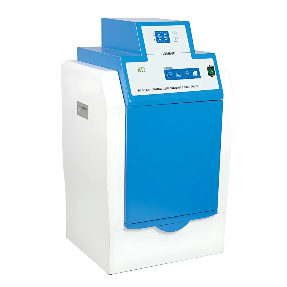
Junyi JY04S-3C Gel Imaging System
| Brand | Junyi |
|---|---|
| Origin | Beijing, China |
| Manufacturer Type | Authorized Distributor |
| Region of Manufacture | Domestic (China) |
| Model | JY04S-3C |
| Instrument Type | Standard Gel Imaging System |
| CCD Resolution | 1.4 MP (1280 × 1024) |
| Bit Depth | 10-bit |
| Dynamic Range | 0–4.8 OD |
| CCD Sensor Size | 2/3" |
| Detection Sensitivity | <20 pg double-stranded DNA stained with ethidium bromide (EB) |
| Signal-to-Noise Ratio | ≥56 dB |
| Lens | F/1.2, 8–48 mm 6× zoom lens |
| UV Transillumination Area (W×L) | 250 × 200 mm |
| Visible Light Transillumination Area (W×L) | 250 × 210 mm |
| UV Transillumination Wavelength | 302 nm (8 W) |
| UV Reflectance Wavelengths | 254 nm & 365 nm (11 W each) |
| Dimensions (L×W×H) | 470 × 405 × 820 mm |
| Net Weight | 29.0 kg |
Overview
The Junyi JY04S-3C Gel Imaging System is a dedicated digital imaging platform engineered for post-electrophoresis visualization, documentation, and quantitative analysis of nucleic acid and protein gels. It operates on the principle of fluorescence excitation and emission capture—utilizing high-intensity, narrow-band UV transillumination (302 nm) and reflectance (254 nm / 365 nm), combined with precisely matched optical filtering (590 nm emission cutoff), to isolate target band signals from background noise. The system integrates a scientific-grade, low-light-sensitive monochrome CCD sensor (2/3″ format, 10-bit digitization, 1.4 megapixel resolution) with a motorized 6× f/1.2 zoom lens assembly, enabling consistent spatial calibration across magnification levels and high-fidelity signal acquisition even at sub-nanogram DNA mass thresholds (<20 pg dsDNA with EB staining). Designed for routine molecular biology laboratories, it delivers reproducible, traceable image capture under controlled darkroom conditions while maintaining full compliance with biosafety requirements for UV exposure containment.
Key Features
- Imported high-sensitivity, low-noise CCD sensor with 10-bit analog-to-digital conversion and ≥56 dB signal-to-noise ratio for reliable detection of faint bands.
- Motorized 6× variable-focus zoom lens (8–48 mm, f/1.2) supporting precise framing, focus adjustment, and consistent pixel-to-mm calibration across magnifications.
- Integrated dual-mode illumination: 302 nm UV transilluminator (8 W) with 250 × 200 mm active area; dual-wavelength UV reflectance (254 nm & 365 nm, 11 W each) for surface fluorescence applications.
- Optimized optical path featuring multi-layer anti-reflective coated 590 nm bandpass emission filter to suppress stray light and enhance contrast in ethidium bromide-stained gels.
- Intelligent dark chamber with programmable safety logic: automatic UV shutoff upon door opening, user-activated UV override for gel excision, and timed UV auto-off (programmable delay).
- Modular mechanical design including drawer-style gel tray with integrated centering LED guide, removable UV-blocking cutting hood, and interchangeable white-light reflectance panel for Coomassie- or SYBR-safe-stained gels.
- Full remote control via USB 2.0 or RS-232 interface—including lens zoom/focus/aperture, UV/white-light activation, and exposure time—enabling integration into automated lab workflows.
Sample Compatibility & Compliance
The JY04S-3C accommodates standard vertical and horizontal electrophoresis gels (up to 25 cm width), including agarose, polyacrylamide (PAGE), SDS-PAGE, and native gels stained with ethidium bromide, SYBR® Gold, GelRed®, Coomassie Brilliant Blue, or silver nitrate. Its optical configuration meets ISO 15195:2019 requirements for analytical instrument validation in clinical molecular pathology labs when used with documented SOPs. UV lamp output and shielding conform to IEC 62471 (Photobiological Safety of Lamps) Category 2 (Moderate Risk) classification, and all safety interlocks satisfy EN 61010-1:2010 for laboratory electrical equipment. While not FDA 21 CFR Part 11–certified out-of-the-box, the system supports audit-trail-capable third-party imaging software compliant with GLP/GMP documentation standards.
Software & Data Management
The system operates with Junyi’s proprietary GelAnalysis Pro™ software (Windows-compatible), providing real-time preview, exposure optimization, background subtraction, lane/band detection, molecular weight estimation, and relative quantification (integrated density analysis). All image metadata—including exposure time, lens position, lamp status, date/time stamp, and operator ID—are embedded in TIFF or PNG exports. Raw image files retain unprocessed 10-bit linear data for reanalysis. Export formats include PDF reports with annotated lanes, Excel-compatible quantification tables, and publication-ready RGB TIFFs. Software supports batch processing, template-based report generation, and direct network printing. For regulated environments, optional validated software modules provide electronic signature support, change control logs, and 21 CFR Part 11–compliant audit trails.
Applications
- Qualitative and semi-quantitative analysis of PCR products, restriction digests, and cloning constructs.
- DNA ladder calibration and fragment sizing with ±3% accuracy across 50 bp–10 kb range (dependent on gel matrix and staining method).
- Protein expression profiling via SDS-PAGE with Coomassie or silver stain densitometry.
- Verification of CRISPR editing efficiency through T7E1 or Surveyor nuclease assays.
- Documentation and archiving of electrophoretic results for internal QC records, regulatory submissions (e.g., ISO 17025 accreditation), and collaborative data sharing.
- Teaching laboratory use—supporting student-led experiments with intuitive interface, real-time visualization, and export functionality for lab notebooks.
FAQ
What is the minimum detectable DNA mass using ethidium bromide staining?
The system reliably detects double-stranded DNA bands containing less than 20 pg when stained with ethidium bromide under standard 302 nm UV excitation.
Can the JY04S-3C be used with SYBR Safe or other non-EB dyes?
Yes—the 302 nm transilluminator and 590 nm emission filter are compatible with SYBR Safe, GelRed, and similar green-fluorescent nucleic acid dyes; optimal exposure times may vary.
Is the UV transilluminator uniform across the entire gel area?
Illuminance uniformity across the 250 × 200 mm active area is maintained within ±12% (measured at 1 cm grid intervals), ensuring consistent band intensity for quantitative comparisons.
Does the system support time-lapse or multi-exposure bracketing?
Yes—GelAnalysis Pro™ allows automated exposure series acquisition (e.g., 0.1–60 s increments) to identify optimal integration time without saturation.
How is calibration maintained over time?
The motorized lens includes positional encoders for repeatable zoom/focus settings; users can save and recall calibrated configurations per gel type and staining protocol.


