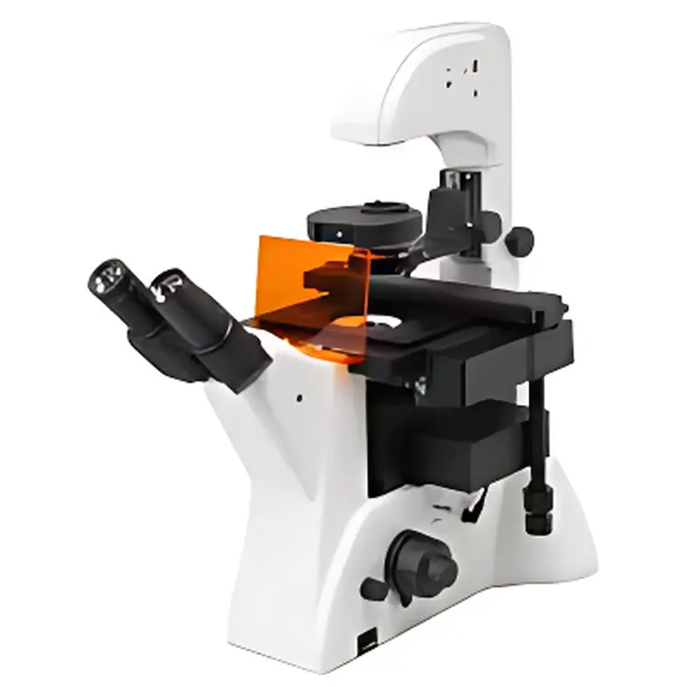
LEI-TECH LK-YG900 Inverted Fluorescence Microscope
| Brand | LEI-TECH |
|---|---|
| Origin | Tianjin, China |
| Manufacturer Type | Original Equipment Manufacturer (OEM) |
| Product Category | Domestic |
| Model | LK-YG900 |
| Instrument Type | Inverted Fluorescence Microscope |
| Excitation Source | High-Stability LED |
| Medical Device Classification | Non-Medical |
| Microscope Class | Conventional Fluorescence Microscope |
| Eyepieces | Widefield Plan Eyepieces PL10X/22mm |
| Objective Lenses | 10X, 20X, 40X (Infinity-Corrected) |
| Fluorescence Filter Sets | DAPI (UV), GFP (B), Cy3 (G) |
| Transmitted Light Source | 6V 30W Halogen Lamp |
| Control Mode | Manual |
| Focusing Mechanism | Coaxial Coarse/Fine Focus with Slip-Resistance Adjustment |
| Condenser | Long Working Distance (55 mm) Condenser |
| Observation Head | Ergonomic Binocular Hinged Head, 45° Inclination, Interpupillary Distance 53–75 mm |
| Stage Travel Range | 77 mm (Y) × 134.5 mm (X), Removable Mechanical Stage Scale |
| Camera Interface | C-mount, 20 MP Sensor (5440 × 3648 pixels) |
| Optical System | Infinity-Corrected Chromatic Aberration-Corrected (UISC) Design |
| Total Magnification Range | 100×–400× |
| Imaging Capabilities | Real-Time Z-Stack Depth Fusion, Multi-Field Mosaic Acquisition, HDR Image Processing (DHR), Onboard Measurement Tools |
Overview
The LEI-TECH LK-YG900 Inverted Fluorescence Microscope is engineered for high-fidelity live-cell and tissue culture observation in academic, industrial, and preclinical research laboratories. Its inverted optical architecture positions the objective lenses beneath the specimen stage—enabling direct access to cell culture vessels including Petri dishes, multi-well plates, and flasks—while maintaining stable thermal and mechanical conditions during extended imaging sessions. The microscope employs an infinity-corrected optical pathway (UISC design), which decouples magnification from tube length, allowing modular integration of auxiliary components such as phase contrast sliders, fluorescence filter cubes, and digital imaging interfaces without compromising resolution or contrast. This architecture ensures consistent wavefront fidelity across all magnifications and illumination modes. The system supports three primary observation modalities: brightfield (BF), phase contrast (PH), and epifluorescence—with dedicated LED excitation sources and bandpass filter sets optimized for DAPI (350–400 nm excitation), GFP (450–490 nm), and Cy3 (530–560 nm). Unlike finite-conjugate systems, the UISC platform minimizes chromatic aberration and field curvature, delivering sharp, flat-field images suitable for quantitative morphometric analysis and longitudinal time-lapse studies.
Key Features
- Infinity-corrected optical system (UISC) with apochromatic-grade color correction and minimal spherical distortion across the full field of view
- Dual-illumination configuration: Stable 6V/30W halogen lamp for transmitted-light brightfield and phase contrast; high-output, thermally regulated LED modules for fluorescence excitation (UV/B/G channels)
- Manual coaxial focusing mechanism with dual-speed fine adjustment (1 µm graduation) and anti-slip tension control—designed for reproducible Z-positioning during serial sectioning or time-lapse acquisition
- Long-working-distance condenser (55 mm) compatible with thick-bottomed cultureware and objective working distances up to 5.1 mm (at 40X)
- Ergonomic binocular head with 45° viewing angle, adjustable interpupillary distance (53–75 mm), and dual light-path split (100:0 / 0:100) for simultaneous eyepiece viewing and camera output
- Large mechanical stage with 77 × 134.5 mm travel range, removable vernier scale, and optional stage clips for secure positioning of standard culture formats (e.g., 35 mm dishes, 6–96-well plates)
- Integrated 20-megapixel CMOS imaging module (C-mount interface, 5440 × 3648 resolution) supporting real-time HDR (DHR) processing, pixel-level intensity normalization, and on-device measurement tools (distance, area, angle, profile intensity)
Sample Compatibility & Compliance
The LK-YG900 accommodates a broad spectrum of biological specimens under physiologically relevant conditions: adherent mammalian cells (HeLa, NIH/3T3, CHO), suspension cultures (Jurkat, primary lymphocytes), organoids, zebrafish embryos, and thin tissue explants. Its inverted geometry eliminates the need for coverslip mounting—reducing shear stress and enabling continuous perfusion or gas exchange during imaging. The stage and condenser are calibrated for use with standard tissue culture plastic (0.17 mm thickness tolerance) and glass-bottom dishes. While not classified as a medical device per FDA 21 CFR Part 809 or ISO 13485, the instrument meets IEC 61000-6-3 (EMC emissions) and IEC 61010-1 (safety for laboratory equipment) standards. Its optical performance aligns with ISO 8578 (microscope resolution test protocols) and ASTM E2812 (fluorescence filter characterization guidelines), ensuring traceable repeatability in comparative assays.
Software & Data Management
The bundled acquisition software provides native support for TIFF, JPEG2000, and OME-TIFF export formats—ensuring compatibility with open-source analysis platforms (Fiji/ImageJ, QuPath, CellProfiler). It implements audit-trail logging compliant with GLP documentation requirements: timestamped metadata capture includes exposure time, gain, LED intensity, objective ID, filter position, and stage coordinates. All image operations—including HDR fusion, Z-stack projection (max-intensity, average, standard deviation), and multi-field mosaic stitching—are performed in real time without post-processing latency. Raw data files retain embedded EXIF tags for instrument configuration, facilitating cross-platform validation and regulatory submission readiness (e.g., FDA eCTD modules).
Applications
- Live-cell tracking of fluorescently labeled organelles (mitochondria, lysosomes, ER) over 24–72 hr periods
- Quantitative assessment of transfection efficiency using GFP/RFP reporter constructs
- Morphological screening of drug-induced cytotoxicity in 96-well plate assays
- Phase contrast-based confluence monitoring in bioreactor seed train workflows
- Co-localization analysis of nuclear (DAPI) and cytoplasmic (Cy3) markers in fixed monolayers
- Time-lapse documentation of wound-healing migration in scratch assays
FAQ
Is the LK-YG900 suitable for long-term time-lapse imaging?
Yes—the LED excitation source generates negligible heat (<1.2 W thermal load at full intensity), and the inverted design minimizes mechanical perturbation to cultured specimens. Optional environmental chamber integration (not included) enables CO₂ and temperature control.
Can third-party objectives be mounted on this microscope?
Yes—the nosepiece uses standard RMS (Royal Microscopical Society) threading and supports any infinity-corrected objective with 45 mm parfocal distance and 200 mm tube lens specification.
Does the system support automated focus drift correction?
No—focus stabilization requires external hardware (e.g., piezo Z-stage or hardware-based focus lock). The manual coarse/fine focus is optimized for precision but lacks motorized feedback loops.
What fluorescence filter compatibility does the LK-YG900 offer beyond DAPI/GFP/Cy3?
The four-slot filter cube turret accepts standard 25 mm diameter cubes. Users may replace factory-installed sets with custom configurations (e.g., TRITC, Alexa Fluor 647, mCherry) provided they match the LED excitation bandwidth and emission cutoff specifications.
Is remote operation or network connectivity supported?
The acquisition software operates locally via USB 3.0; no built-in Ethernet/WiFi interface is provided. Remote desktop solutions (e.g., VNC, TeamViewer) may be deployed for off-site supervision, though real-time streaming bandwidth exceeds 120 Mbps at full sensor resolution.


