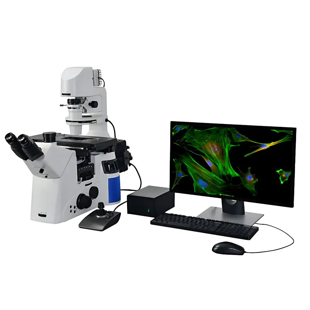
Lei-Tech LK-YG96 Research-Grade Automated Inverted Fluorescence Microscope
| Brand | Lei-Tech |
|---|---|
| Origin | Tianjin, China |
| Manufacturer Type | Direct Manufacturer |
| Instrument Type | Inverted Fluorescence Microscope |
| Model | LK-YG96 |
| Excitation Source | High-Stability LED |
| Observation Modes | Brightfield, Darkfield, Phase Contrast, DIC, Fluorescence |
| Eyepieces | Widefield PL10X/25 mm (adjustable diopter) |
| Objectives | Infinity-Corrected Semi-Apochromatic Plan Phase Contrast Objectives (4X, 10X, 20X, 40X, 60X) |
| Fluorescence Filter Sets | B, G, UV bands with high-transmission, low-autofluorescence interference filters |
| Illumination | Dual LED System (10 W Transmitted + 10 W Reflected) |
| Focus Mechanism | Motorized Coaxial Coarse/Fine Focus with 0.02 µm minimum step resolution and ±0.1 µm repeatability |
| Condenser | Motorized Revolving Condenser (NA 0.55, WD 26 mm), accommodates up to 6 modules (Phase, DIC, BF) |
| Observation Head | Trinocular Tube with Bertrand Lens, 45° inclination, interpupillary adjustment 47–78 mm |
| Stage | Motorized XY Stage (130 × 100 mm travel, 0.1 µm resolution, ±0.5 µm repeatability, max speed 10 mm/s) |
| Objective Turret | Motorized 6-Position Turret with DIC slot and objective protection logic |
| Fluorescence Filter Wheel | Motorized 6-Position Filter Cube Selector |
| Imaging System | 20 MP CMOS Sensor (5440 × 3648 active pixels) |
| Optical Design | Infinity-Corrected Dual-Path UISC (Universal Infinite Space Correction) System |
Overview
The Lei-Tech LK-YG96 is a research-grade automated inverted fluorescence microscope engineered for rigorous live-cell imaging, longitudinal tissue culture observation, and multi-modal quantitative microscopy in academic, pharmaceutical, and biotechnology laboratories. Built upon an infinity-corrected dual-path UISC optical architecture, the system eliminates chromatic and spherical aberrations across all magnifications and modalities—ensuring consistent wavefront fidelity from brightfield through UV-excited fluorescence. Its inverted configuration provides unobstructed access to bottom-mounted specimens in standard Petri dishes, multi-well plates, and perfusion chambers—critical for time-lapse imaging of adherent mammalian cells, organoids, and zebrafish embryos. The integrated LED illumination platform delivers stable, flicker-free excitation (B/G/UV bands) and Köhler-transmitted illumination with color temperature consistency (3700–5000 K), eliminating thermal drift and enabling long-duration acquisitions without phototoxicity escalation.
Key Features
- Motorized multi-axis precision control: Synchronized XY stage (130 × 100 mm travel), Z-focus (0.02 µm step resolution), 6-position objective turret, and 6-slot fluorescence filter wheel—all programmable via unified software interface.
- Dual LED illumination architecture: Independent 10 W transmitted LED (Köhler-optimized) and 10 W reflected LED (spectrally matched to B/G/UV filter sets), each with intensity modulation and TTL triggering support for synchronized camera exposure.
- UISC optical pathway: Proprietary infinite-conjugate design supporting simultaneous phase contrast, DIC, and fluorescence without mechanical realignment; enables seamless modality switching during acquisition protocols.
- Long-working-distance motorized condenser: NA 0.55, WD 26 mm, with six-module electric rotation for rapid interchange of phase annuli, DIC prisms, and BF apertures—fully compatible with thick samples and environmental chambers.
- High-fidelity imaging suite: 20 MP scientific CMOS sensor (5440 × 3648) with on-chip binning, hardware-level HDR capture, real-time Z-stack synthesis, and mosaic tiling (multi-field stitching) with sub-pixel registration accuracy.
- Ergonomic trinocular head: 45° inclined tube with Bertrand lens for Köhler alignment verification, adjustable interpupillary distance (47–78 mm), and dedicated C-mount port for camera coupling with 1×/1.5×/CF intermediate magnification options.
Sample Compatibility & Compliance
The LK-YG96 supports standardized life science specimen formats including 35 mm–150 mm Petri dishes, 6–384-well microplates, chambered coverslips, and custom flow-cell mounts. Its 26 mm working distance condenser and 60X semi-apochromatic objective (WD ≥ 0.15 mm) accommodate live samples under glass-bottom vessels with embedded sensors or gas-permeable membranes. All optical components comply with ISO 8578 (microscope mechanical tolerances) and ISO 10934-1 (optical performance testing). While not classified as a medical device (non-FDA 510(k)/CE IVD), the system meets GLP-compliant documentation requirements: full audit trail logging (user actions, parameter changes, acquisition timestamps), electronic signature support, and metadata-embedded TIFF export compliant with NIH ImageJ/Fiji and OMERO interoperability standards.
Software & Data Management
The proprietary Lei-Tech MicroSuite v4.2 provides a modular, scriptable acquisition environment supporting multi-dimensional acquisition (XYZTλ), autofocus routines (contrast-based and pattern-matching), and batch processing pipelines. Software modules include Dynamic HDR (adaptive pixel-wise gain mapping to suppress saturation while preserving dim signal), Real-Time Extended Depth of Field (EFD) synthesis using focus-stepping algorithms, and Seamless Mosaic Stitching with distortion correction and intensity normalization. Raw image data are saved in TIFF format with embedded EXIF-like metadata (objective ID, filter position, exposure time, LED intensity %, stage coordinates). Export options include OME-TIFF for FAIR data principles compliance, and direct upload to institutional LIMS or cloud repositories (AWS S3, NAS) with configurable encryption (AES-256). Audit logs meet FDA 21 CFR Part 11 requirements for electronic records and signatures when enabled.
Applications
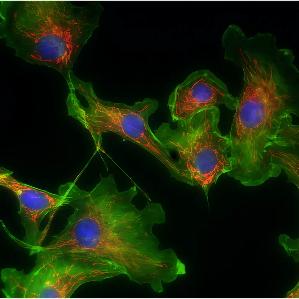
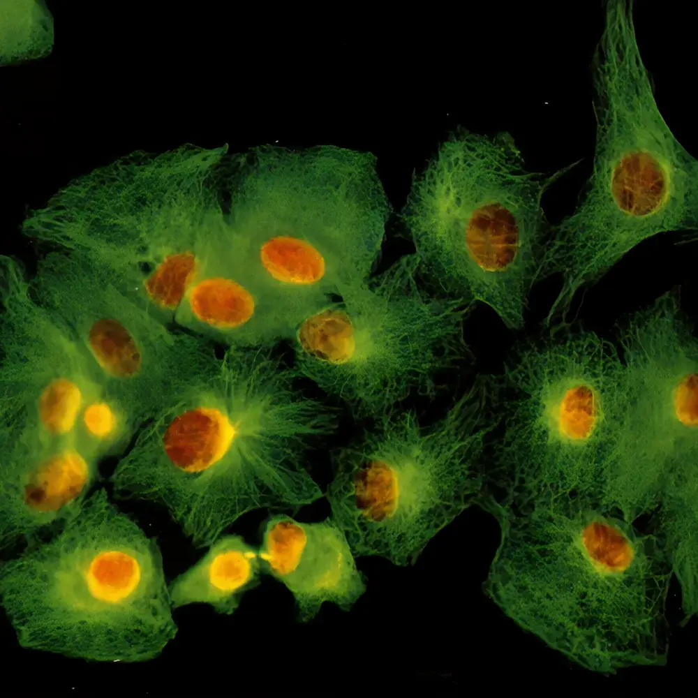
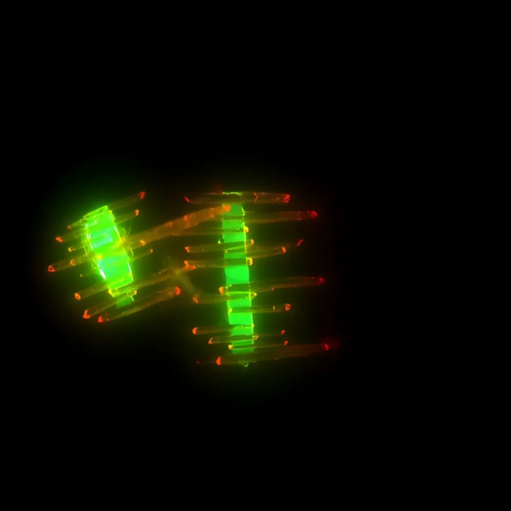
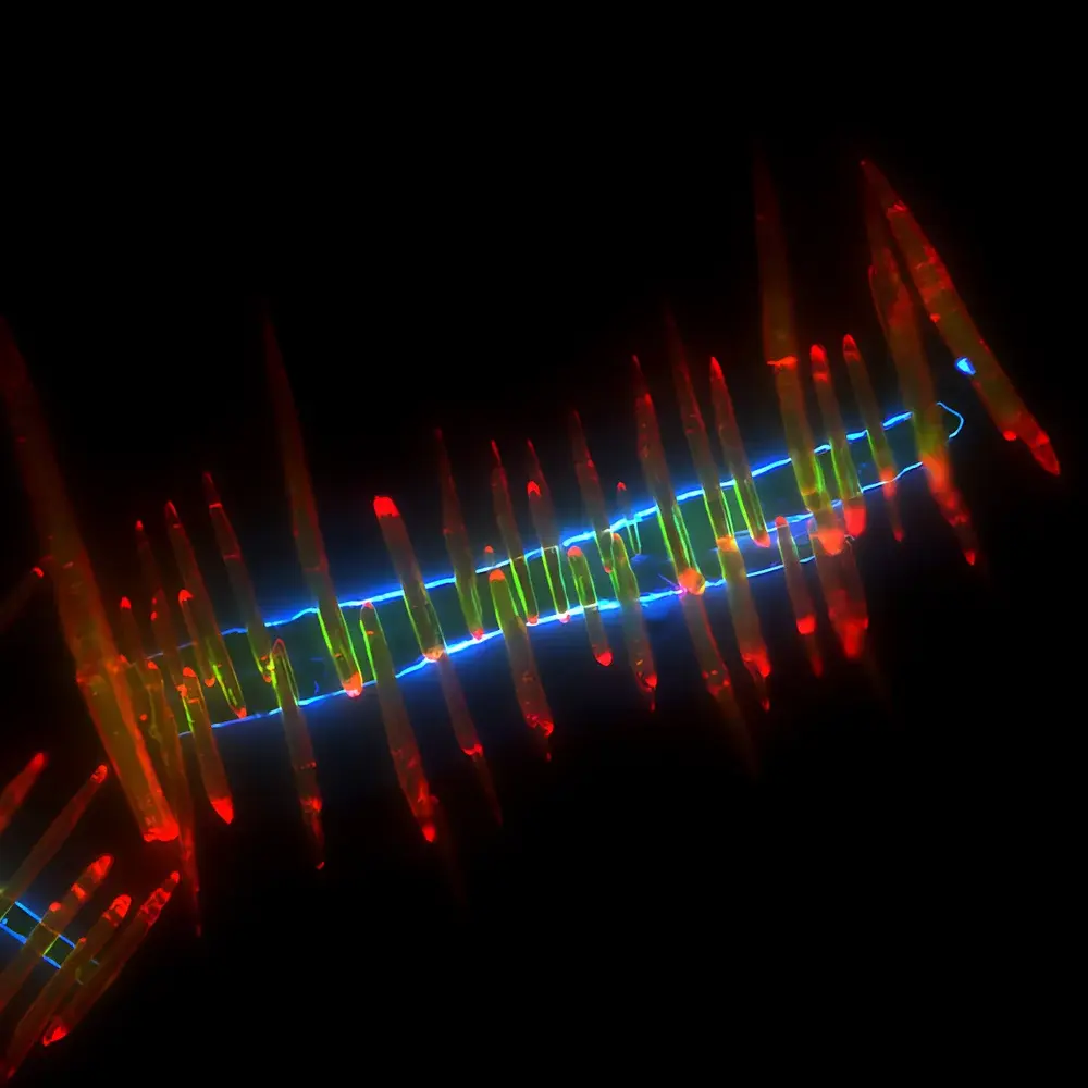
- Live-cell dynamics: Long-term tracking of GFP/RFP-tagged proteins in adherent cultures under controlled CO₂/humidity environments—leveraging low-heat LED excitation and motorized stage for multi-position time-lapse.
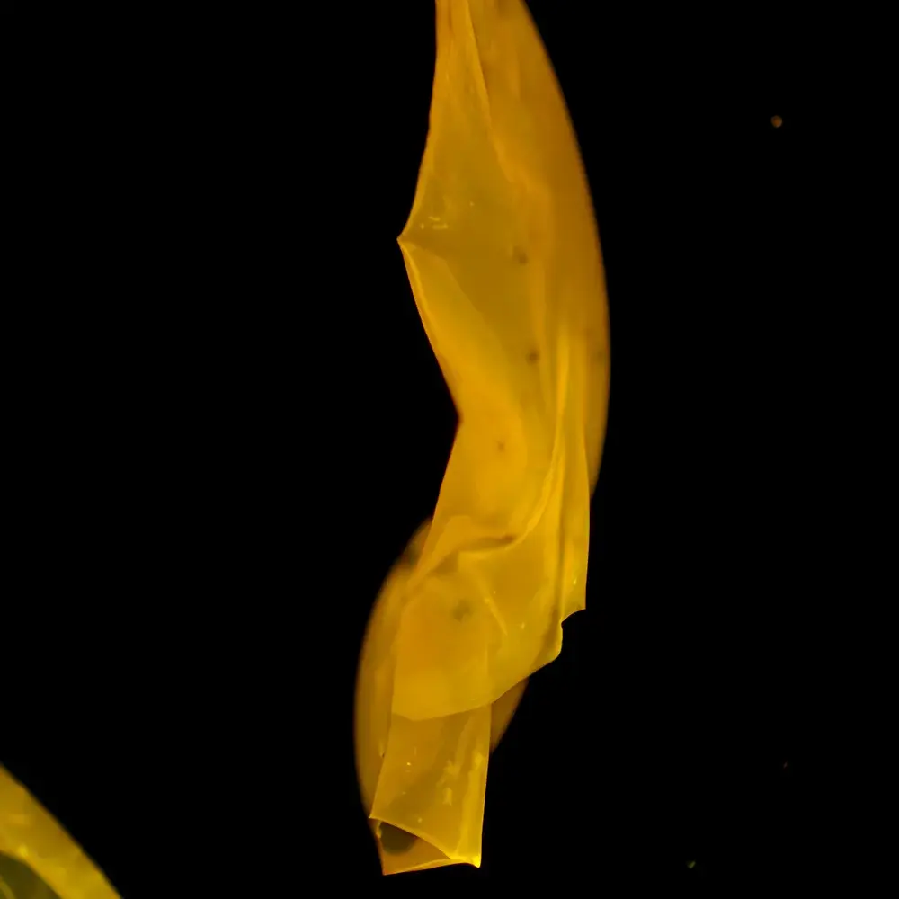
- 3D tissue imaging: Confocal-grade optical sectioning via Z-stack acquisition combined with DIC contrast for label-free structural analysis of spheroids and explants.
- Quantitative fluorescence assays: Multi-channel co-localization (B/G/UV) with spectral unmixing support, background subtraction, and integrated intensity profiling across regions of interest (ROIs).
- High-content screening (HCS): Automated well-to-well scanning in 96-/384-well plates using stage coordinate mapping and adaptive focus correction per well.
- Developmental biology: Embryo imaging in glass-bottom dishes with DIC + fluorescence overlay, supported by thermal-stable optics and vibration-damped baseplate.
FAQ
Is the LK-YG96 compatible with third-party cameras or software platforms?
Yes—the microscope features standard C-mount and USB3.0/PCIe interfaces, and provides SDK documentation for integration with Micro-Manager, Python (PyVISA, OpenCV), and MATLAB via serial/TTL command protocol.
What maintenance is required for the LED light sources?
LED modules are rated for >25,000 hours at nominal output; no lamp replacement or alignment is needed. Annual calibration of intensity uniformity and spectral output is recommended for quantitative workflows.
Does the system support environmental chamber integration?
Yes—stage cutouts and cable routing ports accommodate commercial incubation chambers (e.g., Tokai Hit, OkoLab); the motorized components operate reliably within 20–40°C and 30–80% RH ranges.
Can DIC and fluorescence be used simultaneously?
Yes—the UISC optical path and motorized condenser/objective turret allow concurrent DIC prism insertion and fluorescence filter selection without vignetting or alignment loss.
What warranty and service coverage is provided?
Lei-Tech offers a 24-month comprehensive parts-and-labor warranty, backed by regional service centers across North America, EU, and APAC—with remote diagnostics, on-site installation, and application-specific training included.


