
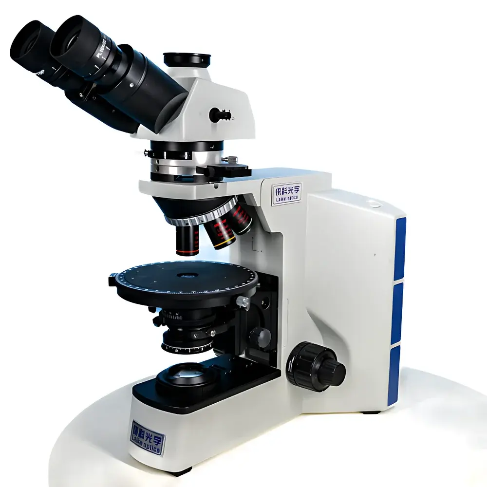
LEI-TECH LK53P Research-Grade Polarizing Microscope for Pharmaceutical Transmitted/Reflected Light Analysis
| Key | Brand: LEI-TECH |
|---|---|
| Model | LK53P |
| Origin | Tianjin, China |
| Manufacturer Type | OEM Manufacturer |
| Optical System | Infinity-Corrected (UISC) |
| Total Magnification | 50×–500× |
| Eyepieces | PL10×/22 mm with reticle |
| Objectives | 5×, 10×, 20×, 50× (achromat, polarized-light optimized) |
| Illumination | Dual-mode (transmitted Köhler + reflected halogen, 12 V/50 W, pre-centered) |
| Polarization Components | Rotatable analyzer (0–360°, 2° scale, ±6′ accuracy), removable λ-plate (551 nm), λ/4-plate (147.3 nm), quartz wedge (I–IV order), built-in Bertrand lens |
| Stage | 360° rotatable metal stage with graphite coating |
| Condenser | Swing-out achromatic condenser (NA 1.2), integrated polarizer with 0°/90°/180°/270° indexing |
| Focus Mechanism | Coaxial coarse/fine focus with tension adjustment and upper limit stop |
| Camera Interface | C-mount, 20 MP sensor (5440 × 3648 effective pixels) |
| Software Features | Real-time Extended Focus Imaging (EFI), Multi-Field Image Assembly (MIA), quantitative measurement, image annotation & calibration |
| Compliance | Designed per ISO 8578 (microscope mechanical standards), compatible with GLP documentation workflows and ASTM E112 / E1181 for grain/phase analysis |
Overview
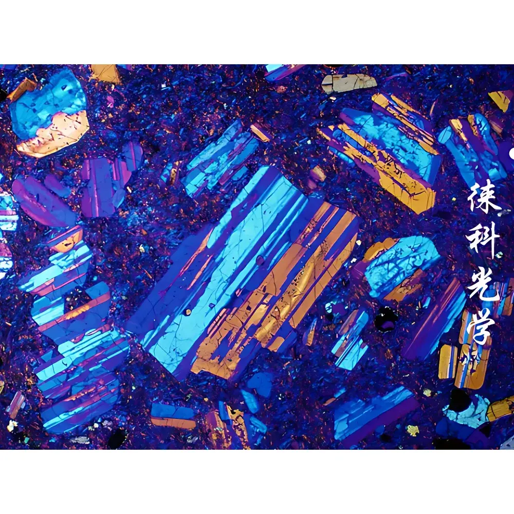
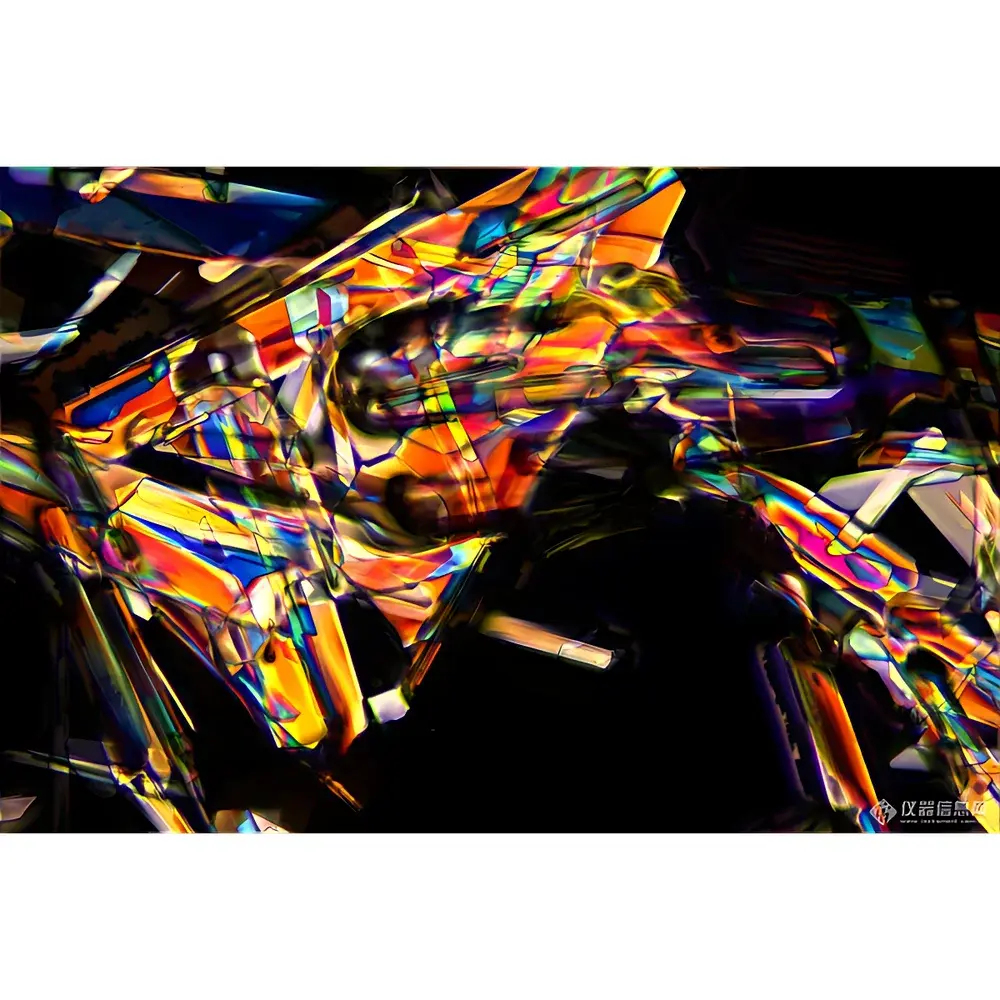
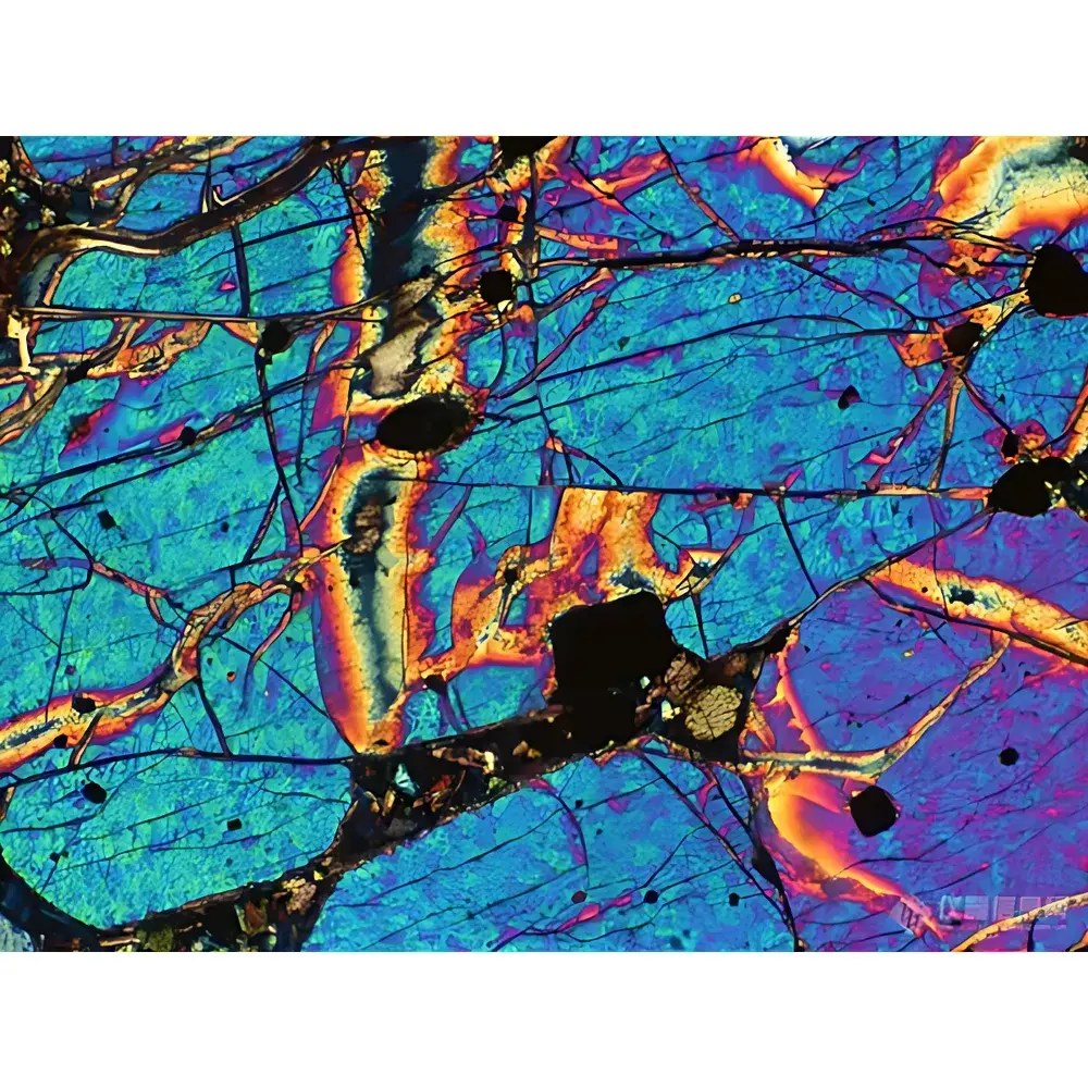
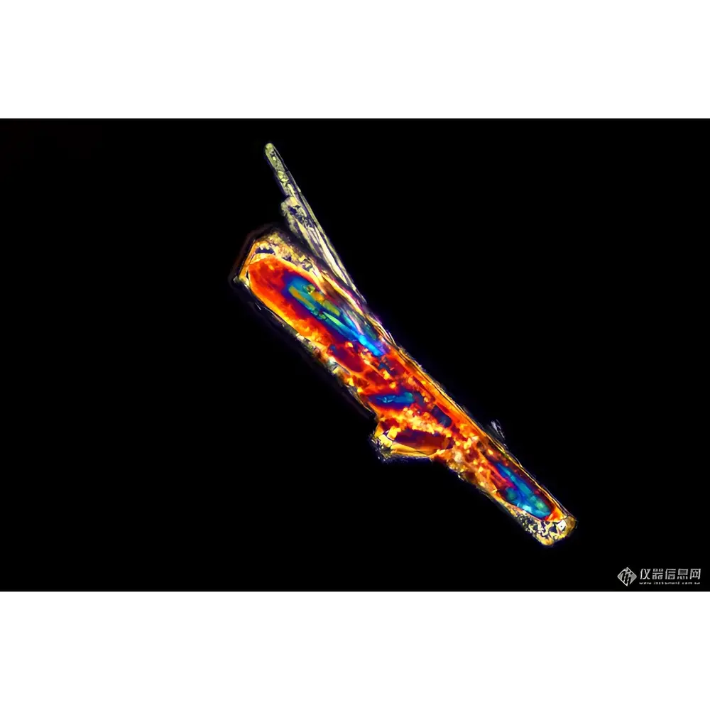
The LEI-TECH LK53P is a research-grade polarizing microscope engineered for high-fidelity birefringence characterization in pharmaceutical solid-state analysis, geological mineralogy, polymer science, and advanced materials R&D. It employs an infinity-corrected optical path (UISC architecture), eliminating chromatic and spherical aberrations across the full magnification range (50×–500×). This design ensures consistent wavefront fidelity—critical for quantitative retardation measurements and interference color interpretation under crossed polars. The system supports both transmitted-light Köhler illumination and incident-light halogen illumination, enabling simultaneous or sequential observation of transparent crystalline domains (e.g., active pharmaceutical ingredients, hydrates, co-crystals) and opaque or reflective samples (e.g., metallurgical phases, coated tablets, composite cross-sections). Its dual-path capability meets USP and ICH Q5A requirements for polymorph identification and phase purity assessment in drug development laboratories.
Key Features
- Infinity-corrected UISC optical system with strain-free objectives and stress-optimized prisms—minimizes artificial birefringence induced by mechanical mounting or thermal gradients.
- Dual-illumination architecture: Transmitted path features a swing-out achromatic condenser (NA 1.2) with integrated indexed polarizer; reflected path includes pre-centered 12 V/50 W halogen lamp, adjustable field and aperture diaphragms, and LBD filter set for optimal contrast in fluorescence-free polarized imaging.
- Rotatable analyzer turret with 2° engraved scale and ±6′ angular repeatability—enables precise extinction angle determination for crystallographic orientation mapping.
- Modular compensation system: Interchangeable λ-plate (551 nm, first-order red), λ/4-plate (147.3 nm), and calibrated quartz wedge (I–IV order)—supports quantitative retardation measurement from sub-nm to >1000 nm ranges per ASTM E1181.
- Ergonomic inverted trinocular head: 30° inclined, interpupillary adjustment (54–75 mm), ±5 D diopter correction per eyepiece, and selectable beam splitting (100:0 or 50:50) for simultaneous visual observation and digital capture.
- Real-time computational imaging suite: Includes Extended Focus Imaging (EFI) for depth-resolved stacking without Z-motor dependency, and Multi-Field Image Assembly (MIA) for seamless wide-area mosaics—both fully integrated into the acquisition software with pixel-level registration and metadata embedding.
Sample Compatibility & Compliance
The LK53P accommodates standard 25 mm and 40 mm diameter thin sections, polished metallographic mounts, pharmaceutical tablet cross-sections, polymer films, and fluid inclusions in epoxy-embedded specimens. Its stage permits full 360° rotation with positive locking—essential for universal stage measurements per ASTM E112 (grain size) and ISO 9042 (crystal habit quantification). All optical components comply with ISO 8578 mechanical tolerances for microscope frame stability and alignment retention. The system’s hardware and software architecture support audit-trail-enabled operation per FDA 21 CFR Part 11 when deployed with validated third-party acquisition platforms. Calibration certificates for analyzer rotation, compensator retardation values, and magnification linearity are available upon request—traceable to NIM (National Institute of Metrology, China) reference standards.
Software & Data Management
The bundled acquisition software provides native support for TIFF-64, JPEG2000, and OME-TIFF export formats—with embedded EXIF and custom metadata fields (polarizer angle, compensator type, exposure time, objective ID, stage coordinates). Measurement tools include linear distance, area, angle, and grain boundary tracing with ISO 23278-compliant stereological sampling protocols. All EFI and MIA datasets retain full Z-stack and mosaic coordinate metadata, enabling downstream reconstruction in open-source platforms such as QuPath or Fiji. Raw image buffers are stored in lossless mode; processed overlays (e.g., isochromatic contour maps) are saved separately to preserve analytical integrity. Exported reports conform to GLP template structures—including operator ID, instrument serial number, timestamped calibration logs, and version-controlled software build identifiers.
Applications
- Pharmaceutical solid-state characterization: Identification of polymorphic forms (e.g., ritonavir Form I vs. II), detection of residual solvents via birefringent inclusion morphology, and assessment of crystallinity distribution in direct-compression tablets.
- Geological thin-section analysis: Quantitative determination of optic sign, 2V angle, and extinction angles in silicate minerals using quartz wedge and accessory plates—aligned with procedures in Deer, Howie & Zussman (1992).
- Polymer phase morphology: Visualization of spherulite growth kinetics, lamellar orientation in polyethylene blends, and stress-induced birefringence in injection-molded components.
- Metallurgical microstructure: Grain boundary delineation in austenitic stainless steels, identification of delta ferrite in duplex alloys, and intermetallic phase mapping in Ni-based superalloys under crossed polars with λ-plate enhancement.
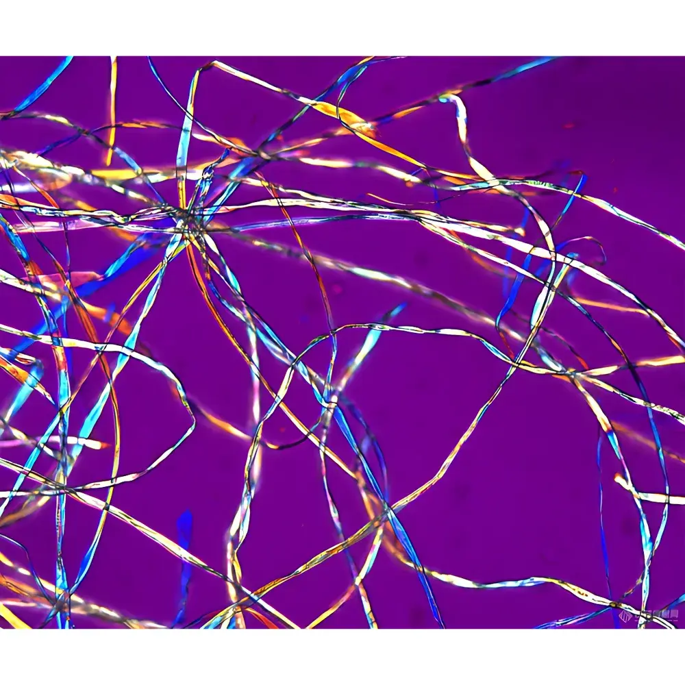
- Forensic fiber analysis: Discrimination of synthetic fibers (e.g., PET vs. nylon 6,6) based on characteristic interference colors and dispersion staining behavior.
FAQ
Does the LK53P meet international calibration traceability requirements for regulated labs?
Yes—optical and mechanical calibrations can be performed using NIM-traceable artifacts. Optional IQ/OQ documentation packages are available for GMP-aligned validation.
Can the system interface with third-party image analysis platforms like HALO or Visiopharm?
Yes—via standardized TIFF/OME-TIFF export with embedded spatial metadata and objective magnification tags.
Is the halogen lamp compatible with automated intensity stabilization during time-lapse birefringence monitoring?
The lamp power supply supports external TTL-triggered dimming; closed-loop intensity feedback requires optional photometric sensor integration.
What is the maximum usable field of view for MIA stitching at 500× magnification?
At 500× (50× objective + 10× eyepiece), the stitched mosaic covers up to 1.2 mm × 0.8 mm with sub-pixel registration accuracy (±0.3 µm RMS).
Are replacement λ-plates certified for retardation value and uniformity?
All compensators ship with individual test reports specifying measured retardation across the central 12 mm aperture, verified using a calibrated photoelastic comparator per ISO 11477.


