Leica Cell DIVE Ultra-Multiplex Tissue Imaging & Spatial Biomarker Analysis System
| Brand | Leica |
|---|---|
| Origin | Germany |
| Manufacturer Type | Authorized Distributor |
| Origin Category | Imported |
| Model | Cell DIVE |
| Pricing | Available Upon Request |
Overview
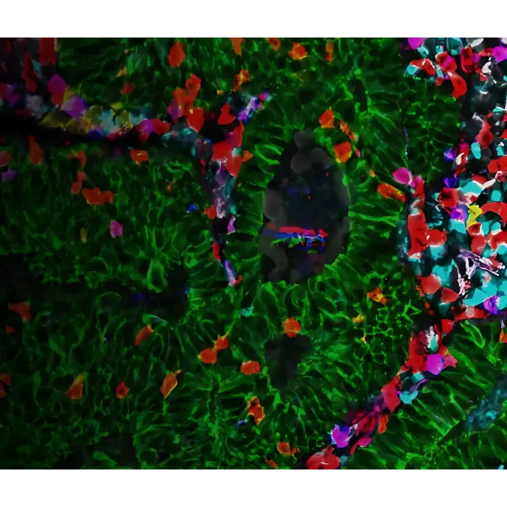
The Leica Cell DIVE Ultra-Multiplex Tissue Imaging & Spatial Biomarker Analysis System is a fully integrated, cyclic immunofluorescence (CycIF)-based platform engineered for high-plex, spatially resolved protein profiling in formalin-fixed paraffin-embedded (FFPE) and frozen tissue sections. Operating on the principle of iterative antibody labeling, imaging, and gentle fluorophore inactivation—without antigen epitope stripping or tissue degradation—the system enables quantitative, subcellular-resolution mapping of up to 60+ protein biomarkers from a single tissue section. Unlike conventional multiplex immunofluorescence (mIF) platforms limited to 6–8 markers per run, Cell DIVE leverages proprietary photochemical bleaching and automated registration algorithms to ensure pixel-perfect alignment across cycles, preserving native tissue architecture while delivering robust, reproducible spatial phenotyping data at single-cell resolution. Designed specifically for translational oncology, immuno-oncology, and spatial biology research, Cell DIVE bridges the gap between bulk omics and functional histopathology by generating comprehensive spatial interaction networks among immune cells, stromal components, and malignant populations.
Key Features
- Ultra-high-plex capability: Sequential detection of ≥60 protein targets per tissue section via cyclic immunofluorescence with minimal signal carryover
- Single-cell spatial resolution: Submicron (<0.3 µm/pixel) imaging supported by Leica’s high-NA objective optics and precision motorized stage
- Gentle, non-destructive cycling: Photochemical fluorophore inactivation preserves antigen integrity and tissue morphology across >30 labeling-imaging-bleach cycles
- Automated workflow integration: Fully coordinated hardware control (microscope, camera, environmental chamber, fluidics) with real-time focus stabilization and drift correction
- Validated antibody panel: Access to >120 rigorously tested, cross-validated primary antibodies—including human immune checkpoint, lineage, functional, and structural markers—with documented lot-to-lot consistency
- End-to-end calibration: On-instrument auto-calibration of illumination uniformity, chromatic aberration, and z-stack depth ensures quantitative comparability across sessions and instruments
Sample Compatibility & Compliance
Cell DIVE supports standard FFPE and OCT-embedded frozen sections (4–10 µm thickness) mounted on charged glass slides. The protocol is compatible with routine H&E staining, prior IHC, and most common antigen retrieval methods (e.g., citrate-EDTA pH 6.0/9.0). All reagents—including proprietary bleaching buffers and blocking solutions—are supplied as CE-IVD marked kits where applicable and manufactured under ISO 13485 quality management systems. The platform meets essential requirements for GLP-compliant spatial biomarker studies and supports audit-ready documentation aligned with FDA 21 CFR Part 11 principles, including electronic signatures, user access logs, and immutable instrument metadata capture per imaging cycle.
Software & Data Management
Acquisition and analysis are managed through Leica Application Suite X (LAS X) Spatial Edition, featuring modular workflows for cycle planning, multi-channel registration, cell segmentation (using deep learning-based nuclei/cytoplasm classifiers), and neighborhood analysis. Raw image stacks (OME-TIFF compliant) are automatically annotated with MIAME/MINSEQE-compliant metadata. Quantitative outputs include marker intensity distributions, cellular co-expression matrices, spatial proximity metrics (e.g., nearest-neighbor distances, Ripley’s K-function), and interactive UMAP/t-SNE visualizations. Data export supports FCS, CSV, and HDF5 formats for downstream integration with R/Bioconductor (e.g., SPATA, histoCAT) or Python-based spatial transcriptomics pipelines (e.g., squidpy, scanpy). Audit trails record all processing parameters, user actions, and software version history per dataset.
Applications
- Spatial tumor microenvironment (TME) characterization in immuno-oncology clinical trial biopsies
- Identification of predictive immune cell spatial signatures associated with response to PD-1/PD-L1 inhibitors
- Mapping of stromal-immune crosstalk in fibrotic or inflammatory disease models
- Validation of spatially resolved targets identified via spatial transcriptomics (e.g., Visium, Xenium)
- Development of AI-powered digital pathology classifiers trained on multiplexed spatial phenotypes
- Longitudinal biomarker tracking in preclinical PDX or GEMM cohorts
FAQ
How many cycles can be performed on a single tissue section without compromising morphology?
Typical workflows execute 30–45 labeling-imaging-bleach cycles with no observable loss of tissue integrity or nuclear detail, as confirmed by H&E re-staining and electron microscopy validation.
Is antibody stripping required between cycles?
No. Cell DIVE uses non-covalent, light-mediated fluorophore inactivation—eliminating harsh chemical stripping that damages epitopes and causes delamination.
Can Cell DIVE data be integrated with single-cell RNA-seq datasets?
Yes. The platform generates cell-level feature vectors (protein expression + spatial coordinates) compatible with multimodal alignment tools such as Seurat v5’s integration framework and Tangram.
What level of technical support is provided for assay development?
Leica offers application scientist-led experimental design consultation, panel optimization services, and on-site training—including hands-on protocol transfer and QC benchmarking against reference standards.
Does the system support multiplexed DNA/RNA co-detection?
Cell DIVE is optimized for protein detection; however, optional hybridization modules (e.g., RNAscope-compatible protocols) are available under collaborative development agreements with Advanced Cell Diagnostics and Bio-Techne.


