Leica Dendrite Expert Metallurgical Microscope System
| Brand | Leica |
|---|---|
| Origin | Germany |
| Model | Dendrite Expert |
| Configuration | Upright |
| Total Magnification | 1000× |
| Eyepiece | 10× |
| Objective | 100× |
| Compliance | ISO 9001, ASTM E112, ISO 643, USP <788>, GLP/GMP-ready software architecture |
Overview
The Leica Dendrite Expert is a purpose-built metallurgical microscopy and image analysis system engineered for quantitative dendritic microstructure characterization in cast metals, alloys, and solidification research. It operates on the principle of high-resolution brightfield and polarized light imaging coupled with algorithm-driven morphometric segmentation—enabling precise identification, delineation, and measurement of primary and secondary dendrite arm spacing (PDAS/SDAS) in as-cast or heat-treated metallic samples. Designed for laboratories requiring traceable, auditable, and repeatable microstructural quantification—particularly in aerospace-grade aluminum, nickel-based superalloys, titanium, and steel castings—the system integrates optical hardware, calibrated digital imaging, and validated analytical workflows into a single platform compliant with international metallographic standards.
Key Features
- Upright metallurgical microscope configuration optimized for reflected-light observation of polished, etched, or unetched metallic specimens—equipped with Leica’s Plan Apochromat objectives delivering diffraction-limited resolution and chromatic fidelity at 100× magnification.
- Dedicated dendrite recognition engine utilizing adaptive thresholding, edge-enhanced segmentation, and branch-point detection algorithms to distinguish dendritic arms from interdendritic eutectic phases or porosity.
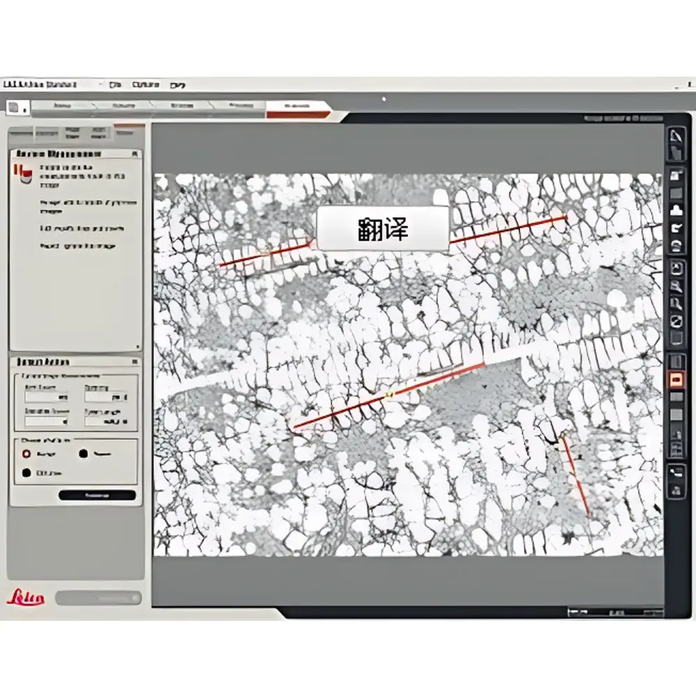
- Two complementary measurement modes: semi-automatic (operator-initiated seed point selection followed by autonomous arm tracing and spacing calculation) and manual (freehand dendrite skeletonization with user-defined arm count and length annotation).
- Real-time overlay of measured parameters—including arm spacing distribution histograms, dendrite centerline maps, and arm count annotations—directly onto the original high-fidelity image, enabling immediate visual verification and metrological traceability.
- Hardware synchronization between Leica DM series upright microscopes, Leica DFC digital cameras (with 5.0 MP–20.0 MP sensor options), and motorized stage controllers ensures pixel-to-physical-unit calibration stability across magnifications and acquisition sessions.
Sample Compatibility & Compliance
The system supports standard metallographic specimen formats (25 mm–50 mm diameter, up to 30 mm thickness) mounted in thermosetting resins or conductive carbon mounts. Compatible with common etchants including Kroll’s reagent (Ti), Keller’s reagent (Al), and Nital (Fe/C alloys). All measurements adhere to ASTM E112 (grain size), ISO 643 (steel microstructure), and ASTM E1382 (quantitative metallography). Software audit trails—including user ID, timestamp, calibration log, and parameter versioning—are recorded per analysis session to support GLP and GMP environments. Exported datasets retain full metadata for FDA 21 CFR Part 11 compliance when paired with Leica Application Suite X (LAS X) Enterprise Edition.
Software & Data Management
Powered by Leica Application Suite X (LAS X) Dendrite Module—a CE-marked, IEC 62304 Class B medical device software variant—the system provides configurable report templates exportable directly to Microsoft Excel (.xlsx) with embedded formulas, dynamic charts (e.g., SDAS frequency distributions, cumulative probability plots), and raw coordinate data (X/Y positions of arm intersections, inter-arm distances in µm). All image processing steps—including illumination normalization, background subtraction, and noise suppression—are non-destructive and fully reversible. Raw TIFF and proprietary .lif files preserve bit-depth integrity (12-bit or 16-bit) for retrospective reanalysis. Data storage follows hierarchical folder structures aligned with ISO/IEC 17025 laboratory documentation requirements.
Applications
- Quality control of investment-cast turbine blades and airfoils where SDAS correlates directly with mechanical strength and creep resistance.
- Process optimization in directional solidification and additive manufacturing (e.g., laser powder bed fusion), where dendrite arm spacing serves as a key indicator of local cooling rate and thermal gradient.
- Research into solidification kinetics, including nucleation undercooling, solute partitioning, and constitutional supercooling modeling validation.
- Failure analysis of hot-tearing or microporosity formation linked to dendritic coherency point deviation.
- Interlaboratory round-robin studies requiring standardized reporting frameworks aligned with ISO/TC 17/SC 1 metallurgical testing protocols.
FAQ
What standards does the Leica Dendrite Expert comply with for metallurgical analysis?
It supports measurement traceability to ASTM E112, ISO 643, ISO 13007, and EN 10365—validated via Leica’s factory calibration certificates and documented uncertainty budgets for pixel-to-µm conversion at each objective magnification.
Can the system be integrated with existing Leica microscope platforms?
Yes—Dendrite Expert software is compatible with Leica DM2700 M, DM4 M, DM6 M, and DM2500 M upright metallurgical microscopes equipped with motorized Z-focus, encoded turret, and LAS X-compatible camera interfaces.
Is Excel-based reporting customizable for internal QA templates?
All report fields—including headers, units, statistical descriptors (mean, std dev, P90), and chart types—are editable via XML-based template definitions; no macro programming or external scripting is required.
How is measurement reproducibility ensured across operators?
Through role-based access control, mandatory calibration checks before analysis, and automated logging of all user-modifiable parameters—ensuring identical processing pipelines regardless of operator experience level.
Does the system support batch processing of multiple images?
Yes—LAS X Batch Processor enables queued analysis of multi-field-of-view datasets, with parallel computation across CPU cores and automatic consolidation of results into unified Excel workbooks with sample-level summary statistics.


