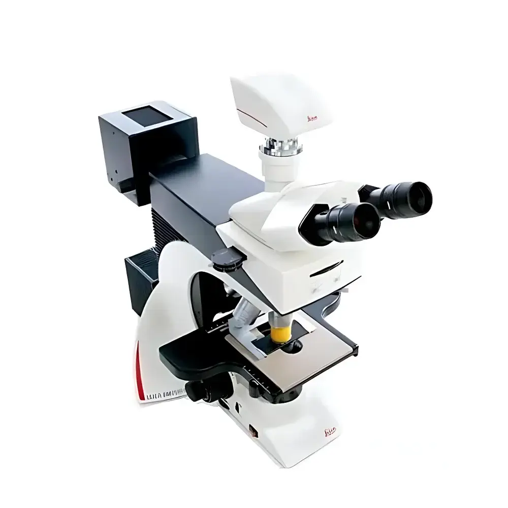
Leica DM2500 Biological Microscope
| Origin | Germany |
|---|---|
| Manufacturer Type | Authorized Distributor |
| Origin Category | Imported |
| Model | DM2500 |
| Pricing | Upon Request |
Overview
The Leica DM2500 is a high-performance upright biological microscope engineered for precision routine diagnostics and advanced life science research. Designed around a robust, modular optical platform, it employs Köhler illumination with a 12 V / 100 W halogen lamp to deliver uniform, stable, and glare-free transmitted-light illumination across all contrast modes—including brightfield, darkfield, phase contrast, polarization, differential interference contrast (DIC), and fluorescence. Its optical architecture complies with international standards for clinical microscopy (e.g., ISO 8578:2017 for microscopes used in medical laboratories) and supports GLP- and GMP-aligned workflows through traceable calibration pathways and documented system configuration. The instrument’s mechanical stability, thermal management of the illumination path, and vibration-damped base ensure high reproducibility in long-duration observations—critical for histopathology slide screening, hematological morphology assessment, and live-cell imaging preparation.
Key Features
- Modular upright stand with ergonomic height-adjustable focusing knobs—patented design accommodating users of varying stature and reducing repetitive strain during extended use
- 7-position revolver nosepiece enabling seamless switching between high-NA objectives without mechanical drift or parfocal shift
- Wide-field binocular or trinocular observation tubes compatible with 22–25 mm field number eyepieces; optional tilting tube (up to 30°) for improved operator posture
- DIC-capable optical train featuring strain-free, high-transmission prisms and adjustable Nomarski shear; optimized for unstained, low-contrast biological specimens such as live cells, tissue sections, and microorganisms
- 5-position rotating fluorescence filter turret with precise mechanical indexing and minimal light leakage; compatible with standard Leica filter sets (e.g., GFP, RFP, DAPI, TRITC)
- Integrated aperture diaphragm control, condenser centering mechanism, and slider-based contrast module selection (phase annuli, polarizers, DIC sliders) for intuitive, tool-free operation
- Universal camera port (C-mount and F-mount options) supporting scientific-grade CCD/CMOS sensors; calibrated for quantitative intensity mapping and pixel-accurate image registration
Sample Compatibility & Compliance
The Leica DM2500 accommodates standard glass slides (1 × 3 inches / 26 × 76 mm), Petri dishes (up to 100 mm diameter), and multi-well plates (6–96-well) via optional stage adapters. Its mechanical stage offers X-Y travel of 76 × 52 mm with vernier scales (0.1 mm resolution) and optional motorized upgrade for tiled scanning. All optical components are certified free of lead and cadmium per RoHS Directive 2011/65/EU. The system meets IEC 61000-6-3 (EMC emission) and IEC 61000-6-2 (immunity) standards. For regulated environments, the microscope supports audit-trail-ready documentation when paired with Leica Application Suite (LAS X) under FDA 21 CFR Part 11-compliant configurations.
Software & Data Management
While the DM2500 is fully functional as a standalone optical instrument, it integrates natively with Leica’s LAS X software platform for image acquisition, annotation, measurement (length, area, intensity profiling), and multi-channel fluorescence overlay. LAS X supports DIC vector analysis, Z-stack reconstruction, and time-lapse metadata tagging—including objective ID, exposure settings, and illumination intensity logs. Raw image data is saved in TIFF or Leica’s proprietary LIF format, both compliant with MIAME and OME-TIFF metadata schemas. Export modules enable direct ingestion into LIS/HIS systems via HL7 or DICOM-SR protocols where configured.
Applications
- Hematology and clinical pathology: Morphologic evaluation of peripheral blood smears, bone marrow aspirates, and cytology preparations under brightfield and phase contrast
- Cell biology: Live-cell imaging support via DIC and phase contrast; compatibility with environmental chambers (temperature/humidity/CO₂ control) for short-term incubation studies
- Microbiology: Identification and motility assessment of unstained bacteria, yeast, and protozoa using darkfield and phase contrast
- Plant science: Observation of stomatal movement, root tip mitosis, and vascular tissue structure with polarization and DIC enhancement
- Quality control in biomanufacturing: Verification of cell culture confluency, viability staining (e.g., trypan blue), and contaminant detection in upstream processing
FAQ
Is the Leica DM2500 suitable for fluorescence applications requiring high signal-to-noise ratio?
Yes—the system features high-transmission optics, a dedicated 5-position fluorescence turret with optimized dichroic mirrors and emission filters, and a stabilized 100 W halogen source with IR-cut filtering to minimize thermal noise in long-exposure acquisitions.
Can the DM2500 be upgraded for digital pathology workflows?
Yes—via optional motorized XY stage, Z-drive, and LAS X Slide Scanning module, enabling automated whole-slide imaging at up to 40× magnification with focus map correction and mosaic stitching.
Does the microscope comply with regulatory requirements for diagnostic laboratories?
Yes—it conforms to ISO 15189:2022 clause 5.3.2 (equipment verification) and supports IQ/OQ documentation packages for clinical deployment in CAP- or CLIA-accredited facilities.
What objective series are recommended for DIC on the DM2500?
Leica HC PL Fluotar and HCX PL FLUOTAR objectives (10×–100×, NA ≥ 0.30) with matched DIC prisms and strain-free correction are validated for zero-pixel-shift DIC imaging.
Is service and calibration support available globally?
Yes—Leica Microsystems maintains an ISO/IEC 17025-accredited service network with certified field engineers and traceable calibration standards (NIST-traceable photometric and geometric references).


