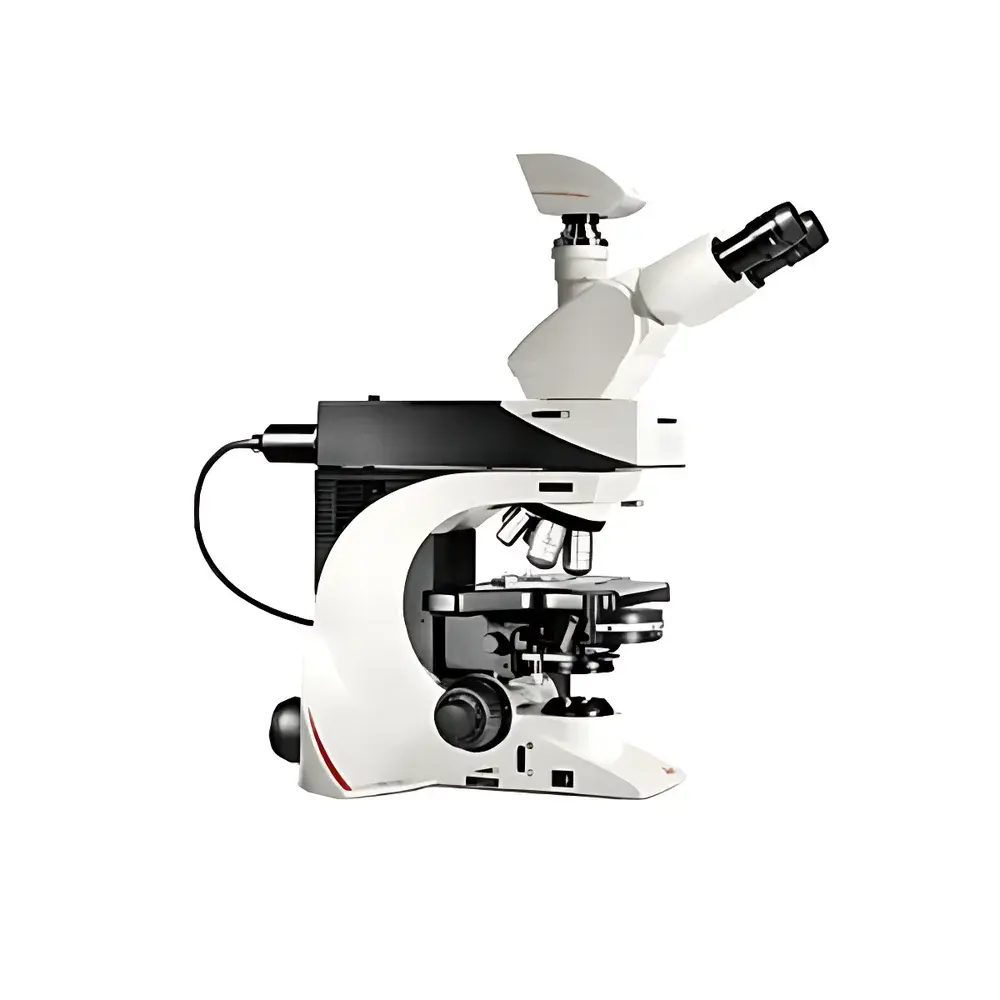
Leica DM2500 Research-Grade Biological Microscope
| Brand | Leica |
|---|---|
| Origin | USA |
| Manufacturer Type | Authorized Distributor |
| Origin Category | Imported |
| Model | Leica DM2500 Research-Grade Biological Microscope |
| Pricing | Upon Request |
Overview
The Leica DM2500 is a research-grade upright biological microscope engineered for high-precision static and dynamic observation in pathology, cytology, hematology, and fundamental life science research. Built upon Leica’s proven infinity-corrected optical architecture, the DM2500 delivers exceptional image fidelity, chromatic and spherical aberration correction, and long-term stability across modalities—including brightfield, darkfield, phase contrast, polarization, fluorescence, and differential interference contrast (DIC). Its modular design supports seamless integration of advanced illumination systems, objective turrets, condensers, and digital imaging components—enabling rigorous, repeatable microscopy under GLP- and GMP-aligned laboratory workflows. The system is optimized for routine diagnostic throughput as well as demanding quantitative fluorescence applications such as co-localization studies, where pixel-level registration integrity is critical.
Key Features
- Infinity-corrected optical pathway with full compatibility for Plan Achromat, Plan Semi-Apochromat, and Plan Apochromat objectives—supporting 4× to 100× magnification with numerical apertures up to 1.40
- 6-position or 7-position motorized or manual nosepiece with precise mechanical indexing and parfocal alignment (≤ ±0.005 mm)
- Dual illumination options: 12 V / 100 W halogen Köhler illumination (30–100 W adjustable) or optional long-life LED illumination with stable color temperature (CCT ≈ 5700 K) and minimal thermal drift
- Rotating 5-position fluorescence filter cube turret with zero-pixel-shift mechanics—ensuring sub-micron spatial registration during multi-channel acquisition
- Ergonomic dual-focus control: coarse/fine focusing with 1 µm fine-step resolution; adjustable torque and focus stop settings; symmetrical left/right hand operation capability
- Ultra-hard ceramic-coated stage (light beige finish) offering chemical resistance, scratch resilience, and enhanced specimen contrast; integrated one-handed specimen clip for rapid slide exchange
- Adjustable inclination (15°–35°) and extendable observation tubes—including ergonomic 15° inclined binocular head and variable-length trinocular port for camera coupling
- Synchronized brightness objectives (HI PLAN SL series): matched luminous flux across 4×, 10×, 20×, and 40× magnifications—eliminating repeated light intensity recalibration during objective switching
- Color-coded aperture diaphragm scale aligned with standard objective color coding (e.g., red = 10×, yellow = 40×), enabling rapid condenser alignment per magnification
Sample Compatibility & Compliance
The Leica DM2500 accommodates standard 1″ × 3″ (25 × 75 mm) glass slides, coverslips (0.13–0.17 mm thickness), and specialized specimen holders for live-cell chambers, metallurgical sections, and polarized thin sections. It complies with IEC 61000-6-3 (EMC emissions) and IEC 61000-6-2 (immunity), and meets essential safety requirements per EN 61010-1 for laboratory electrical equipment. When configured with FDA-registered imaging software (e.g., Leica LAS X Core or LAS X Life Science Edition), the system supports audit trail generation, electronic signatures, and 21 CFR Part 11 compliance for regulated environments. Optional DIC and polarization modules conform to ISO 10934-1 (microscopy terminology) and ASTM E2812 (quantitative phase measurement standards).
Software & Data Management
The DM2500 interfaces natively with Leica’s LAS X platform—a modular, workflow-driven imaging suite supporting multi-modal acquisition, spectral unmixing, Z-stack reconstruction, time-lapse registration, and quantitative morphometry. LAS X includes built-in calibration management (objective-specific scaling, illumination flat-field correction), metadata embedding (EXIF + custom tags), and export compatibility with TIFF, OME-TIFF, and ND2 formats. For enterprise integration, LAS X supports DICOM-SR export, LDAP authentication, and secure network deployment via TLS 1.2 encryption. Third-party compatibility includes HALCON, ImageJ/Fiji plugins (via TWAIN/DCAM drivers), and MATLAB-based analysis pipelines using Leica’s SDK.
Applications
- Routine histopathological screening and frozen-section analysis in clinical laboratories
- Fluorescence co-localization and FRET studies requiring pixel-accurate channel overlay
- High-magnification DIC imaging of unstained cellular ultrastructure (e.g., cytoskeletal dynamics, organelle motility)
- Quantitative phase contrast analysis of live adherent or suspension cells without fixation or staining
- Polarized light examination of birefringent structures (e.g., collagen fibers, starch granules, mineral crystals)
- Multi-user teaching labs equipped with shared viewing stations and annotation-capable digital output
- Quality control of microfabricated devices and semiconductor wafers using reflected-light modes
FAQ
Is the Leica DM2500 compatible with third-party digital cameras?
Yes—the microscope features standardized C-mount (1×) and F-mount (0.5×) ports, and supports USB 3.0, GigE Vision, and Camera Link interfaces via Leica-certified adapters.
Can the DM2500 be upgraded to support automated stage scanning?
Yes—Leica offers motorized XY stages (e.g., LMS-200) with programmable positioning accuracy of ±1 µm and repeatability of <0.5 µm, fully controllable through LAS X.
Does the zero-pixel-drift fluorescence turret require recalibration after service?
No—the turret’s kinematic mounting and passive thermal compensation eliminate post-service realignment; factory calibration is traceable to NIST standards.
What is the maximum working distance available for high-NA objectives on the DM2500?
The HI PLAN CY objective series provides up to 17 mm working distance at 20×/0.40 NA, optimized for thick specimens and inverted-style sample handling.
How does the DM2500 meet regulatory requirements for diagnostic use?
When paired with validated software, documented IQ/OQ protocols, and traceable calibration certificates, the system fulfills essential criteria for CE-IVD (Class B) and FDA 510(k)-cleared workflows in clinical microscopy.


