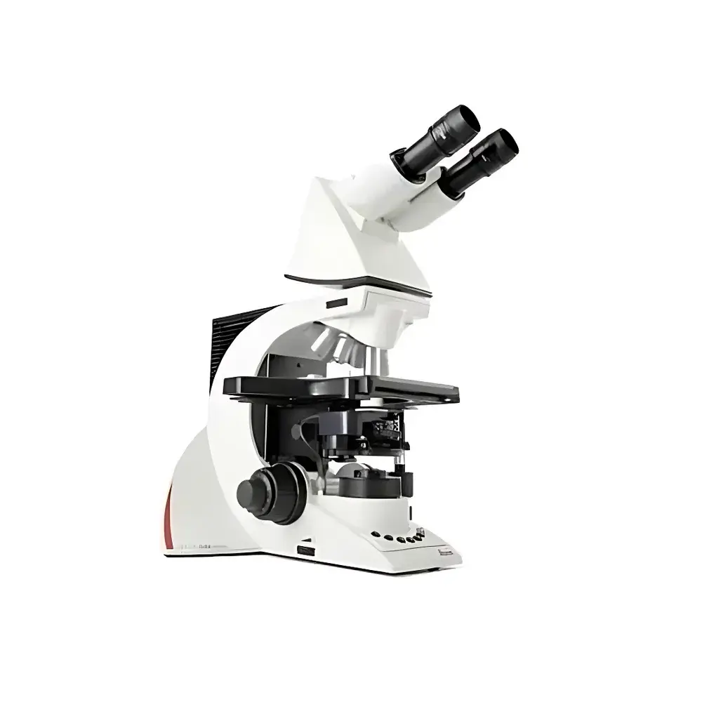
Leica DM3000 Biological Microscope
| Origin | Germany |
|---|---|
| Manufacturer Type | Authorized Distributor |
| Origin Category | Imported |
| Model | DM3000 |
| Pricing | Upon Request |
Overview
The Leica DM3000 is a high-performance, ergonomically engineered biological microscope designed for routine clinical diagnostics and biomedical research applications in pathology, cytology, hematology, and histology laboratories. Built upon Leica Microsystems’ decades-long heritage in optical precision, the DM3000 employs Köhler illumination principles with motorized condenser positioning and dynamic light intensity regulation to ensure consistent, reproducible imaging across magnifications. Its modular architecture supports both upright brightfield observation and optional fluorescence imaging (with compatible filter cubes), making it suitable for standardized diagnostic workflows compliant with ISO 15189, CLIA, and CAP-accredited laboratory environments. The system’s core optical path delivers diffraction-limited resolution across its full suite of HI PLAN apochromatic objectives (4× to 100× oil), with chromatic and spherical aberration correction optimized for high-contrast, low-noise transmission imaging at all magnifications.
Key Features
- Motorized 6-position objective turret with sub-second switching between user-defined objectives — programmable via front-panel buttons or optional footswitch
- Intelligent condenser head (0.90 NA) that automatically positions itself to match each objective’s focal plane and numerical aperture, ensuring optimal Köhler alignment without manual adjustment
- Auto-illuminance control: halogen lamp intensity dynamically adjusts in real time based on selected objective, eliminating manual brightness recalibration during multi-magnification examinations
- Ergonomic focus knob with patented height-adjustable mechanism (DE10340721) — independently adjustable for operator hand size and seated posture
- Symmetrical X-Y stage controls and coaxial focusing for distortion-free, intuitive manipulation; ceramic-coated mechanical stage ensures long-term wear resistance and smooth translation
- Modular trinocular head (HC L1T 4/5/7) compatible with digital camera integration and live image capture; includes daylight filter (32 mm) and dual 10×/22 mm field eyepieces
- Quick-change slide holder enabling one-handed specimen loading; integrated light ring set for phase contrast compatibility (UCA S1)
- Thermal focus compensation system minimizes drift during extended observation sessions, critical for time-sensitive cytological assessments
Sample Compatibility & Compliance
The Leica DM3000 accommodates standard 1″ × 3″ glass slides (up to 1.2 mm thickness) and accommodates coverslips from 0.13–0.17 mm thickness across all objectives. Its optical design complies with ISO 8036-1 (microscope objective standards) and DIN EN 61000-6-3 (EMC emission requirements). When configured with certified digital imaging modules and audit-trail-enabled software (e.g., Leica Application Suite – LAS X Core), the system supports FDA 21 CFR Part 11 compliance for electronic records and signatures in regulated clinical labs. All mechanical components meet RoHS and REACH directives; lamp housing conforms to IEC 61000-4-2 ESD immunity standards.
Software & Data Management
While the base DM3000 operates as a standalone optical instrument, it integrates natively with Leica’s LAS X platform via the HC L1T phototube interface. LAS X Core provides synchronized image acquisition, annotation, measurement (cell counting, area quantification, linear distance), and DIC/phase contrast enhancement algorithms. For pathology workflows, optional LAS X Pathology Edition enables DICOM-SR structured reporting, WSI-compatible tiling, and integration with PACS/LIS systems via HL7 and DICOM protocols. Audit trail logging, user access levels, and electronic signature support align with GLP/GMP documentation requirements. Image metadata (objective ID, magnification, illumination settings, timestamp) is embedded in TIFF and JPEG2000 exports per ISO/IEC 23008-12.
Applications
The DM3000 serves as a primary diagnostic tool in accredited clinical laboratories performing Pap smear analysis, peripheral blood film review, bone marrow aspirate evaluation, and frozen section assessment. Its rapid objective switching and auto-illumination significantly reduce turnaround time in high-volume cytology screening. In academic and pharmaceutical research, the system supports live-cell observation (with environmental chamber compatibility), immunohistochemistry interpretation, and morphometric analysis of tissue sections. Optional fluorescence capability—when paired with Leica’s GFP/RFP/TRITC filter sets—enables basic co-localization studies in cell line characterization and transfection validation.
FAQ
Is the Leica DM3000 compatible with digital camera systems?
Yes — the trinocular phototube (HC L1T 4/5/7) supports C-mount and F-mount adapters for third-party and Leica-certified cameras, with native driver support in LAS X.
Does the DM3000 meet regulatory requirements for clinical diagnostics?
When deployed with validated LAS X software and documented SOPs, the system fulfills essential elements of ISO 15189:2022 and CLIA ’88 for microscopy-based testing.
Can the focus height adjustment be locked after calibration?
Yes — the patented focus knob mechanism includes a mechanical locking collar to prevent unintended repositioning during daily use.
What maintenance is required for the motorized condenser head?
No scheduled maintenance is required; the stepper-motor-driven condenser uses sealed bearings and requires only periodic dust removal using compressed air and lens-grade cleaning tools.
Is thermal focus compensation active during all magnifications?
Yes — the bi-metallic focus drift compensation system operates continuously across the full 4×–100× range, particularly stabilizing high-magnification oil-immersion observations lasting >15 minutes.


