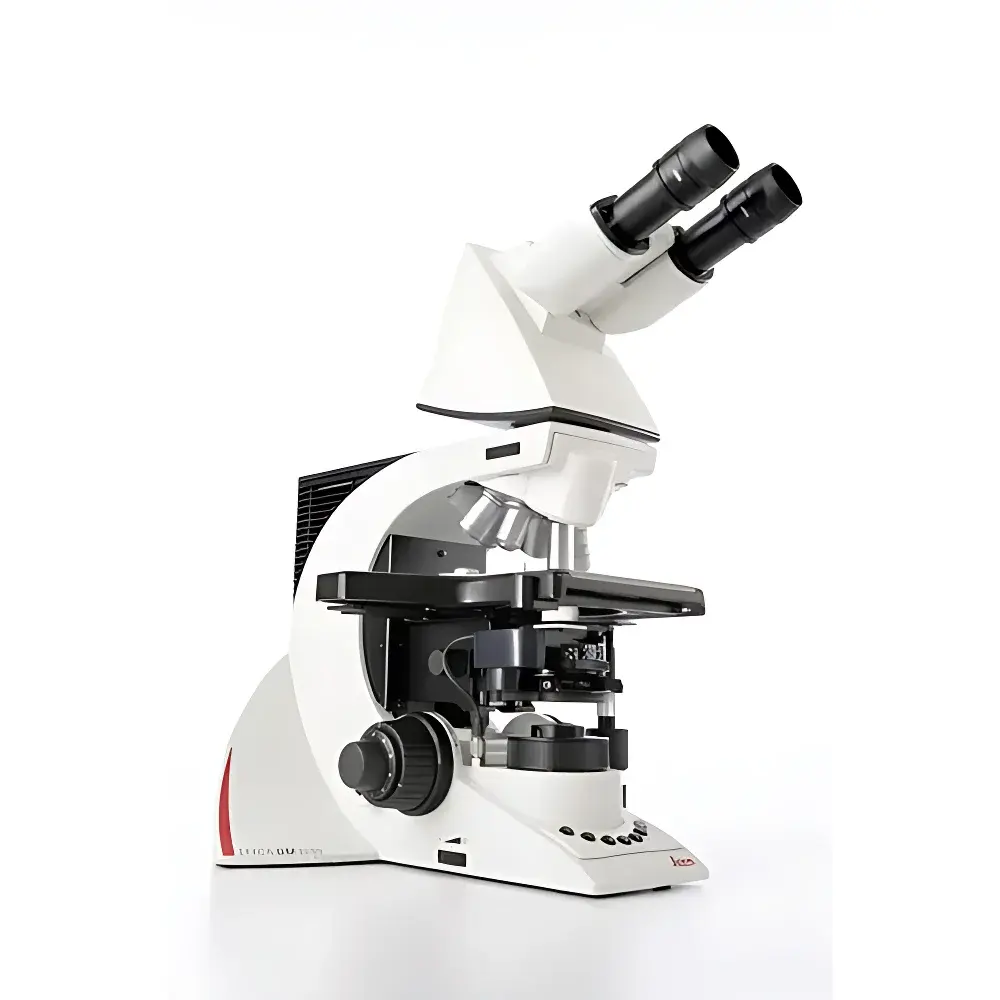
Leica DM3000 LED Upright Biological Microscope
| Brand | Leica |
|---|---|
| Origin | Shanghai, China |
| Manufacturer Type | Authorized Distributor |
| Product Category | Domestic (China-assembled) |
| Model | DM3000 LED |
| Price Range | USD 14,000 – 28,000 |
| Instrument Type | Upright Microscope |
| Illumination | LED |
| Observation Modes | Brightfield, Phase Contrast, Darkfield, Polarization, Fluorescence |
Overview
The Leica DM3000 LED is an upright biological microscope engineered for precision, reproducibility, and ergonomic sustainability in routine and advanced life science laboratories. Built upon Leica Microsystems’ decades of optical expertise, the DM3000 LED employs high-NA achromatic and semi-plan apochromatic objective optics, coupled with a stable Köhler-optimized LED illumination system delivering consistent color temperature (5700 K), flicker-free output, and >25,000-hour lifetime. Its optical path supports simultaneous dual-output imaging—via trinocular port and integrated C-mount—enabling concurrent visual observation and digital documentation without compromise. Designed for clinical pathology, hematology, cytology, and academic cell biology workflows, the system adheres to ISO 10993 biocompatibility standards for user-contact components and complies with IEC 61000-6-3 (EMC) and IEC 60601-1 (medical electrical equipment safety) where applicable.
Key Features
- Motorized nosepiece with six-position turret, enabling programmable, silent, and backlash-free objective switching via front-panel buttons or optional footswitch—reducing manual handling and mechanical drift.
- Automated condenser control: The swing-out top lens of the Abbe condenser disengages automatically when objectives ≤10× are selected, eliminating manual repositioning and maintaining optimal aperture alignment.
- Intelligent LED intensity regulation: Light output dynamically scales with magnification; preset illumination levels per objective are stored and recalled, minimizing phototoxicity and observer eye strain during extended sessions.
- Full ergonomic customization: Adjustable tube inclination (0°–35°), height-adjustable focus knobs (±40 mm range), and symmetric X/Y stage controls reduce upper-body torque and promote neutral posture per ISO 9241-5 ergonomics guidelines.
- Modular stage configuration: Left/right-handed operation is supported via reversible stage control levers; focus knob rear buttons are programmable for image capture, contrast mode toggle, or illumination preset recall.
- Thermally stabilized LED housing ensures minimal thermal drift (<0.1°C/hour at ambient 23°C), preserving specimen integrity during time-lapse observation.
Sample Compatibility & Compliance
The DM3000 LED accommodates standard glass slides (1 × 3 inches), petri dishes (up to 100 mm diameter), and multi-well plates (6–96-well) using optional stage adapters. It supports both transmitted-light techniques—including brightfield, phase contrast (with 10×, 20×, 40× PH objectives), darkfield (dry condenser NA 1.2–1.4), polarization (with rotatable analyzer and strain-free objectives), and fluorescence (with filter cubes for DAPI, FITC, TRITC, and optional LED excitation modules). All optical components meet Leica’s internal QC standards for wavefront error <λ/8 RMS and lateral chromatic aberration <0.5 µm across visible spectrum (400–700 nm). The system is compatible with GLP/GMP documentation workflows through optional Leica Application Suite (LAS X) software with 21 CFR Part 11 audit trail and electronic signature modules.
Software & Data Management
While the DM3000 LED operates as a standalone instrument, it integrates seamlessly with Leica LAS X Core and LAS X Live software platforms via USB 3.0 or Ethernet. LAS X supports real-time Z-stack acquisition, multi-channel fluorescence overlay, measurement annotation (area, length, particle count), and DIC/phase quantification algorithms compliant with ASTM E2921-13 (standard guide for quantitative microscopy). Image metadata—including objective ID, exposure time, LED intensity, condenser position, and user-defined tags—is embedded in TIFF and OME-TIFF formats. Export options include CSV for statistical analysis and DICOM-SR for PACS integration in clinical pathology environments.
Applications
The DM3000 LED serves as a primary platform for diagnostic histopathology (H&E, PAS, Masson’s trichrome evaluation), peripheral blood smear analysis (WBC differential, platelet morphology), urine sediment examination, microbiological identification (Gram staining, acid-fast bacilli), and live-cell observation (unfixed, unstained preparations using phase contrast). Its robust mechanical stage and vibration-damped base support extended-duration imaging in teaching labs and QA/QC settings where regulatory traceability and inter-operator consistency are required per ISO/IEC 17025 clause 7.2.2.
FAQ
Does the DM3000 LED support fluorescence imaging out-of-the-box?
Yes—when equipped with appropriate filter cubes and optional high-power LED excitation modules (e.g., 365 nm, 470 nm, 555 nm), the system delivers stable, low-heat fluorescence excitation suitable for routine epifluorescence applications.
Can the microscope be configured for left-handed users?
Yes—the mechanical stage controls, focus knob orientation, and programmable rear buttons can be fully mirrored or reassigned to accommodate left-hand dominance without hardware modification.
Is the LED illumination intensity calibrated and traceable?
LED output is factory-calibrated against NIST-traceable photometric standards; intensity values are reported in mW/mm² at the specimen plane and logged with each acquired image in LAS X.
What service and calibration options are available internationally?
Leica-certified service centers in North America, EMEA, and APAC provide annual optical alignment verification, LED output certification, and mechanical recalibration per ISO 10110-5, with documented certificates issued upon completion.


