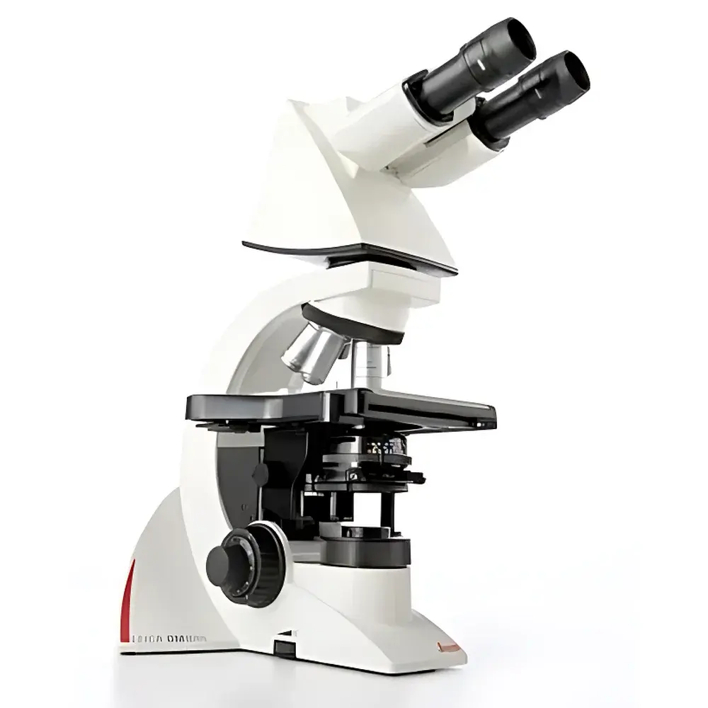
Leica DM3000 Upright Semi-Automatic Fluorescence Microscope (LED-Illuminated)
| Brand | Leica |
|---|---|
| Origin | Shanghai, China |
| Manufacturer Type | Authorized Distributor |
| Product Category | Domestic (China-assembled) |
| Model | DM3000 |
| Price Range | USD 14,000 – 35,000 |
| Instrument Type | Upright Fluorescence Microscope |
| Excitation Source | High-Stability LED |
Overview
The Leica DM3000 Upright Semi-Automatic Fluorescence Microscope is an ergonomically engineered optical platform designed for routine and advanced light microscopy in clinical, diagnostic, and research-oriented life science laboratories. Built upon Leica Microsystems’ legacy of precision optics and modular mechanical architecture, the DM3000 employs Köhler illumination principles with a stable, long-life LED excitation source—eliminating mercury lamp hazards and thermal drift while ensuring consistent intensity across fluorescence channels (DAPI, FITC, TRITC). Its upright configuration supports standard glass slide-based specimen observation, making it especially suited for cytology, hematology, histopathology, and routine immunofluorescence screening. Unlike fully automated systems, the DM3000 retains manual control over critical optical parameters—including focus, stage movement, and filter cube selection—while integrating semi-automated features such as brightness-synchronized illumination and preset turret positions—enabling reproducible workflows without compromising operator engagement or system transparency.
Key Features
- Ergonomic, user-adaptable design including patented height-adjustable coarse/fine focus knobs, tilt-adjustable binocular tube (0–30°), and ambidextrous coaxial stage controls with variable travel length—reducing musculoskeletal strain during extended use.
- LED-based fluorescence illumination system delivering >10,000 hours of stable output, minimal heat emission, and instant on/off switching—fully compliant with ISO 10993 biocompatibility and IEC 62471 photobiological safety standards.
- Modular optical train supporting interchangeable filter cubes (standard 25 mm format), optional phase contrast sliders, and DIC compatibility via add-on components—ensuring flexibility across staining modalities.
- High-stability mechanical stage with vernier scale, dual-axis mechanical drive, and optional motorized XY positioning interface for future integration with image acquisition software.
- Optimized optical path featuring Leica’s HI PLAN achromat objectives (4×–100×, NA 0.10–1.25), offering flat-field correction, high contrast, and chromatic aberration control—validated per ISO 8578 for resolution and MTF performance.
- Compact footprint (base dimensions: 290 × 340 mm) with stackable module architecture—facilitating lab space optimization and compliance with CLIA-certified laboratory spatial requirements.
Sample Compatibility & Compliance
The DM3000 accommodates standard 1″ × 3″ microscope slides and 24 × 50 mm coverslips, with optional extended working distance (ELWD) objectives supporting thick specimens up to 2.5 mm in height—ideal for live-cell imaging in chambered slides or tissue sections mounted in aqueous media. All optical components are certified to ISO 9001:2015 manufacturing standards. The system meets essential requirements of IVD Directive 98/79/EC (as applicable to diagnostic microscopy platforms) and supports GLP-compliant documentation when paired with validated digital imaging software. While not FDA 510(k)-cleared as a standalone diagnostic device, its optical performance aligns with ASTM E2877-21 (Standard Guide for Microscopic Examination of Biological Specimens) and USP environmental monitoring protocols.
Software & Data Management
The DM3000 operates natively with Leica Application Suite (LAS) X Core Edition—a CE-marked, FDA 21 CFR Part 11-ready software platform enabling audit trail logging, user access control, and electronic signature capture. LAS X supports time-lapse acquisition, Z-stack reconstruction, multi-channel fluorescence overlay, and metadata embedding (including objective ID, exposure settings, and filter position). Image files are exported in TIFF, JPEG2000, or Leica’s proprietary .lif format—compatible with third-party analysis tools including ImageJ/Fiji, HALO, and Visiopharm. Optional integration with Leica’s DMC6200 camera enables real-time preview at 30 fps (1920 × 1200) with hardware-based white balance and auto-exposure calibration.
Applications
- Clinical cytology: Routine Pap smear evaluation, urine sediment analysis, and fine-needle aspiration (FNA) preparation screening.
- Hematology: Peripheral blood film differential counts, reticulocyte enumeration, and malaria parasite detection using Giemsa or acridine orange staining.
- Pathology support: Frozen section review, immunohistochemistry (IHC) validation, and fluorescent in situ hybridization (FISH) signal enumeration.
- Academic teaching: Student microscopy labs requiring robust, low-maintenance instrumentation with standardized optical performance.
- Quality control: Raw material inspection of biological reagents, cell culture contamination monitoring, and sterility testing documentation.
FAQ
Is the DM3000 manufactured in Germany?
No—the DM3000 model referenced here is assembled in Leica’s Shanghai facility under strict quality oversight by Leica Microsystems AG. Final optical calibration and QC testing adhere to the same specifications as German-manufactured counterparts.
Can the DM3000 be upgraded to full motorization?
Yes—via Leica’s DM3000-M upgrade kit, which adds motorized nosepiece, Z-drive, and stage control compatible with LAS X automation modules.
Does the LED illumination support quantitative fluorescence intensity measurement?
While the DM3000 provides stable relative intensity, absolute quantification requires external calibration using NIST-traceable fluorescent microspheres and a calibrated photometer—recommended for assay development per ISO/IEC 17025 guidelines.
What is the warranty coverage for this instrument?
Standard warranty is 24 months parts-and-labor, extendable to 60 months under Leica’s ServicePlus program—including annual optical alignment verification and preventive maintenance.
Are replacement LED modules field-serviceable?
Yes—LED modules are tool-free replaceable via front-access drawer mechanism; no optical realignment required post-replacement.


