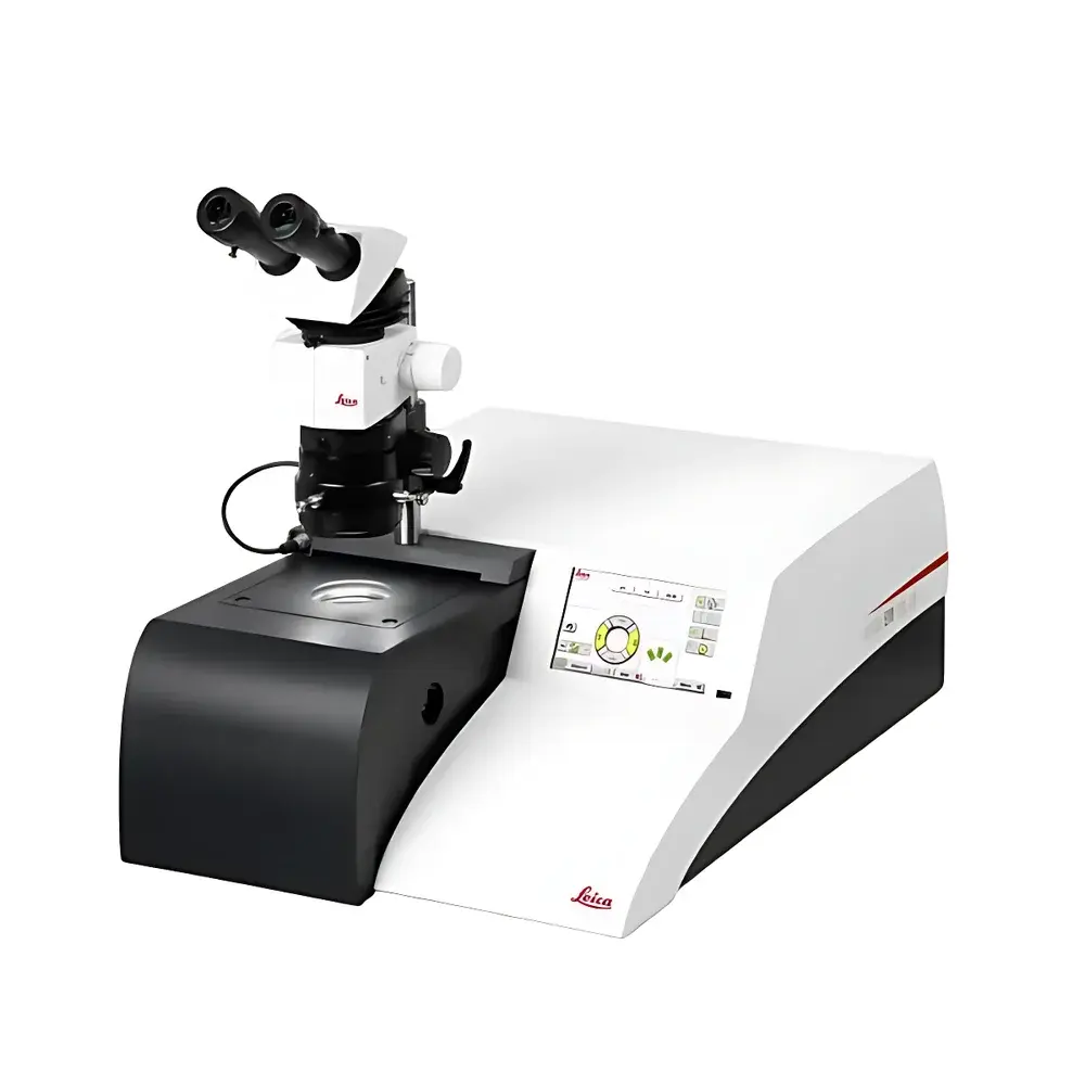
Leica EM TIC 3X Triple-Beam Ion Milling System
| Brand | Leica |
|---|---|
| Origin | Germany |
| Model | Leica EM TIC 3X |
| Sample Chamber Max Size | 50 × 50 × 10 mm |
| Milling Rate (Si) | up to 300 µm/h |
| Cutting Area | >4 × 1 mm |
| Cryo Stage Temp | down to –150 °C |
| Multi-Sample Holder | accommodates up to 3 samples per run |
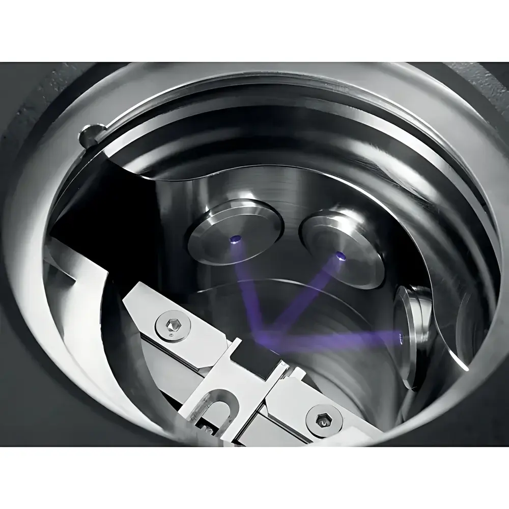
| Ion Beam Configuration | three independently controlled Ar⁺ ion beams |
| Vacuum System | integrated, vibration-isolated turbomolecular pumping |
| Interface | touchscreen control with real-time HD-TV monitoring via built-in camera and LED illumination |
| Data Transfer | USB-based parameter and protocol import/export |
| Contrast Enhancement | in-situ ion beam etching post-milling |
| Compliance | designed for GLP/GMP-aligned lab environments |
Overview
The Leica EM TIC 3X Triple-Beam Ion Milling System is an advanced, fully automated instrument engineered for high-fidelity cross-sectional preparation of heterogeneous, brittle, thermally sensitive, or multi-phase materials—without mechanical deformation, thermal damage, or preferential sputtering artifacts. Unlike conventional single-beam or dual-beam ion mills, the EM TIC 3X employs three independently adjustable argon ion beams operating at precisely controlled angles and energies (typically 1–10 keV), enabling simultaneous broad-area milling, localized fine-tuning, and in-situ contrast enhancement. This tri-beam architecture leverages Couette-type ion flux distribution and optimized beam collimation to achieve uniform material removal across wide fields (>4 mm width) while preserving nanoscale topographic fidelity at the cut surface. Designed for integration into electron microscopy workflows—including scanning electron microscopy (SEM), energy-dispersive X-ray spectroscopy (EDS), wavelength-dispersive spectroscopy (WDS), Auger electron spectroscopy (AES), electron backscatter diffraction (EBSD), and atomic force microscopy (AFM)—the system delivers artifact-free surfaces suitable for quantitative microstructural analysis and phase boundary characterization.
Key Features
- Triple independent ion beam configuration: enables concurrent coarse milling, edge refinement, and surface conditioning without sample repositioning
- Cryogenic sample stage: temperature controllable from –150 °C to +60 °C, critical for stabilizing polymers, biological composites, and low-melting-point alloys during milling
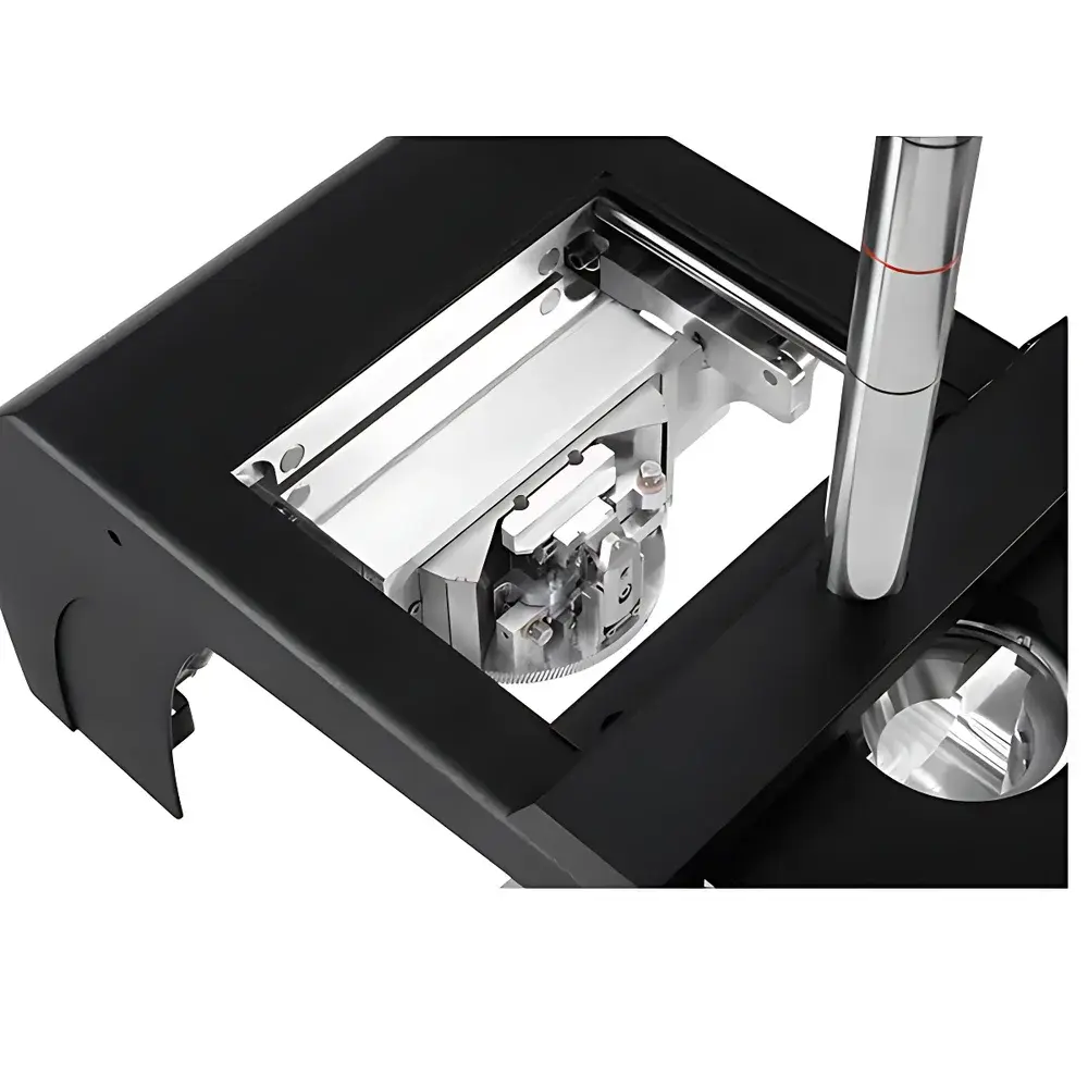
- High-throughput multi-sample holder: accommodates up to three standard 25-mm-diameter or custom-shaped specimens per run, reducing turnaround time and operational overhead
- Vibration-isolated vacuum architecture: integrated turbomolecular pumping system decouples mechanical noise from the observation path, ensuring stable HD-TV imaging and precise endpoint detection
- Real-time visual monitoring: built-in high-definition TV camera with coaxial LED illumination and stereomicroscope-compatible optical interface allows dynamic assessment of milling progression and surface evolution
- In-situ contrast enhancement module: post-milling ion etching performed on the same mount, enhancing topographic contrast of grain boundaries, interfacial layers, and amorphous/crystalline transitions without air exposure or transfer artifacts
- Intuitive touchscreen interface: pre-programmed milling protocols, parameter logging, and USB-based method transfer support reproducible operation across users and shifts
Sample Compatibility & Compliance
The Leica EM TIC 3X supports a broad spectrum of materials—from soft organic matrices (e.g., polymer blends, pharmaceutical tablets, lipid bilayers) to ultra-hard ceramics (e.g., SiC, Al₂O₃), metal-ceramic composites, MEMS devices, geological thin sections, and battery electrode cross-sections. Its non-contact, low-energy ion milling mechanism avoids the plastic deformation common in mechanical polishing and eliminates beam-induced charging or carbon deposition associated with FIB-SEM. The system conforms to laboratory quality management frameworks: audit trails for parameter changes are timestamped and user-logged; all protocols adhere to ASTM E1558 (Standard Guide for Ion Beam Milling of Metallographic Specimens) and ISO 21779:2020 (Electron microscopy — Preparation of cross-sectional specimens by ion beam milling). It is routinely deployed in regulated environments requiring traceability under GLP and GMP guidelines, including failure analysis labs supporting automotive, aerospace, and medical device R&D.
Software & Data Management
Operation is managed via a dedicated embedded Linux-based control suite with a 10.1-inch capacitive touchscreen. Users define milling parameters—including beam energy, incidence angle, current density, dwell time, and cryo-stage setpoint—through intuitive sliders and dropdown menus. All methods are stored as encrypted .xml files and transferred via USB 2.0 for version-controlled archiving. The system logs full operational metadata: vacuum history, beam current stability, stage temperature drift, and real-time image snapshots at user-defined intervals. These logs integrate natively with LIMS platforms through CSV export and support 21 CFR Part 11-compliant electronic signatures when paired with enterprise authentication servers.
Applications
- Failure analysis of solder joints, wire bonds, and die-attach interfaces in semiconductor packaging
- Microstructural quantification of porosity, phase distribution, and interfacial adhesion in solid oxide fuel cells and lithium-ion battery electrodes
- Preparation of lamellar TEM lamellae precursors from bulk specimens prior to FIB lift-out
- Investigation of coating delamination, diffusion zones, and oxidation fronts in thermal barrier coatings
- Correlative microscopy workflows combining AFM topography, EBSD crystallography, and EDS elemental mapping on a single milled surface
- Geological thin-section analysis of metamorphic textures, fluid inclusion networks, and mineral reaction rims
FAQ
What types of samples are incompatible with the Leica EM TIC 3X?
Samples containing volatile components that sublime below –150 °C (e.g., certain hydrated salts or low-boiling solvents) may require pre-stabilization or alternative preparation routes. Highly radioactive or biohazardous specimens must be handled in accordance with institutional radiation safety or biosafety level (BSL) protocols prior to loading.
Can the EM TIC 3X prepare TEM-thin electron-transparent windows?
No—it is not designed for final TEM lamella thinning. However, it excels at producing large-area, flat, and damage-free cross-sections ideal for SEM-based analytics and as substrates for subsequent focused ion beam (FIB) thinning.
Is remote monitoring supported?
While the system does not include native Ethernet-based remote desktop functionality, its USB-exported log files and method archives enable offline review and comparative analysis across instruments in multi-site laboratories.
How is beam alignment verified and maintained?
The system incorporates factory-calibrated beam collimation optics and a reference grid target for periodic verification. No user-accessible alignment tools are required under normal operation; recalibration is performed by certified Leica service engineers using proprietary diagnostic firmware.
Does the cryo-stage require liquid nitrogen refills during operation?
No—the stage uses a closed-cycle Gifford-McMahon cryocooler, eliminating consumables and enabling unattended overnight runs with stable temperature control within ±0.5 °C.


