Leica RUV800 Retinal Upright-Image Viewing System
| Brand | Leica |
|---|---|
| Origin | Germany |
| Manufacturer Type | Authorized Distributor |
| Origin Category | Imported |
| Model | Leica RUV800 |
| Pricing | Upon Request |
Overview
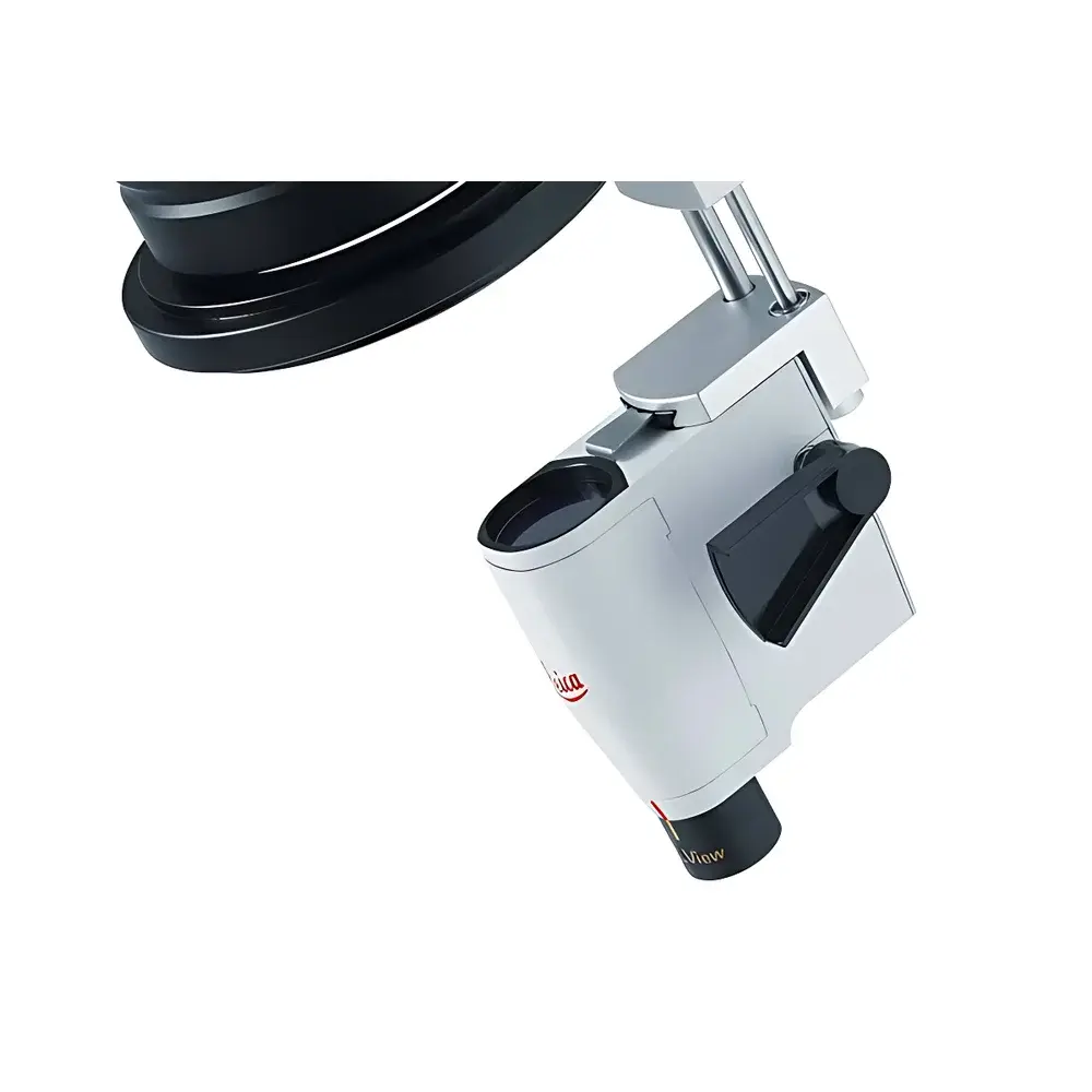
The Leica RUV800 Retinal Upright-Image Viewing System is an optomechanically integrated, non-electronic widefield retinal observation module designed for integration with Leica surgical microscopes. It operates on the principle of optical inversion compensation via a built-in, precision-aligned relay lens system—eliminating the need for external erecting optics or electronic image flipping. Unlike conventional fundus viewing systems that require add-on inverting prisms beneath the binocular tube (which compromise anterior segment ergonomics and optical path length), the RUV800 is mounted directly beneath the objective lens assembly. This architecture preserves the native focal plane alignment and maintains consistent parfocality across both anterior and posterior segment procedures. Engineered for ophthalmic vitreoretinal and combined cataract–vitrectomy workflows, the system delivers real-time, distortion-minimized upright retinal imagery to the surgeon, assistant, and integrated video recording systems—without latency, software dependency, or digital interpolation artifacts.
Key Features
- Optical upright-image generation via integrated relay optics—no electronic processing, no firmware updates, no calibration drift
- Full-field upright imaging across the entire microscope field of view (FOV), including peripheral regions outside the central retinal projection zone
- Quick-swap modular design: RUV800 body remains permanently mounted; only the wide-angle lens (90° or 132°), focusing lever, and silicone sleeve require sterilization between cases
- AgProtect™ nano-silver antimicrobial coating applied to all exposed non-optical surfaces—validated per ISO 22196 for sustained bacterial reduction (>99.9% against S. aureus and E. coli over 24 h)
- Ergonomic height preservation: eliminates vertical extension caused by external erecting prisms, maintaining optimal surgeon posture and reducing neck fatigue during prolonged procedures
- Seamless anterior–posterior modality transition: removal takes <15 seconds; microscope reverts immediately to standard anterior segment configuration compliant with ISO 13485–certified cataract workflow protocols
Sample Compatibility & Compliance
The Leica RUV800 is compatible with Leica M841, M822, and PROve surgical microscope platforms. It supports standard ophthalmic surgical workflows involving phacoemulsification, pars plana vitrectomy, membrane peeling, endolaser photocoagulation, and intraocular lens implantation. The system complies with IEC 60601-1 (medical electrical equipment safety) and IEC 62304 (software lifecycle requirements for embedded optical control logic). Though fully passive (no powered components), its mechanical interface meets EN ISO 15223-1 labeling requirements and carries CE marking under the EU Medical Device Regulation (MDR 2017/745) Class IIa classification. Sterilization validation follows AAMI ST79:2017 guidelines for heat-stable accessories—autoclave cycles up to 134 °C / 3 min are approved for lens housings and focusing levers.
Software & Data Management
As a purely optical, non-digital imaging module, the RUV800 does not incorporate embedded firmware, processors, or data interfaces. It generates no DICOM, JPEG, or video streams natively—instead, it feeds optically corrected upright imagery directly into the microscope’s existing video output pathway (e.g., Leica ICC50 HD or DFC9000 GT cameras). All image capture, annotation, storage, and audit trail functions are managed through the host microscope’s certified software suite, which supports FDA 21 CFR Part 11-compliant user authentication, electronic signatures, and immutable audit logs when deployed in GCP/GLP environments. No additional cybersecurity validation is required for the RUV800 itself due to absence of network connectivity or writable memory.
Applications
- Vitreoretinal surgery requiring panoramic visualization of peripheral retina, lattice degeneration, or retinal breaks
- Combined phacovitrectomy: uninterrupted transition from capsulorhexis to vitreous base shaving without repositioning or recalibration
- Teaching and proctoring: simultaneous upright viewing for primary surgeon, assistant, and OR display monitors—eliminating cognitive load from mental image inversion
- Intraoperative documentation: compatibility with Leica’s certified surgical video archiving systems for retrospective case review and peer-reviewed publication
- Low-vision surgical training: reduces learning curve for residents by preserving natural spatial orientation during instrument manipulation within the eye
FAQ
Does the RUV800 require power, software updates, or routine calibration?
No. It is a fully passive optical module with no electronics, batteries, or firmware. Zero maintenance beyond standard microscope servicing and periodic optical cleaning per Leica Service Bulletin SB-RUV800-03.
Can the RUV800 be used with non-Leica surgical microscopes?
No. Mechanical and optical coupling is proprietary to Leica’s M-series and PROve platform mounting interfaces. Adaptors for third-party systems are neither offered nor validated.
Is the 132° lens compatible with all vitrectomy gauge systems (23G, 25G, 27G)?
Yes. Field-of-view geometry remains consistent across standard transconjunctival trocar configurations; magnification scaling is preserved per Leica Optical Path Specification OPS-RUV800 Rev. 4.2.
How does AgProtect™ coating interact with standard enzymatic or hydrogen-peroxide-based sterilants?
Independent testing (TÜV SÜD Report No. T19-012217) confirms coating integrity retention after 500 autoclave cycles and exposure to Ortho-phthalaldehyde (OPA), glutaraldehyde, and vaporized H₂O₂—no silver ion leaching detected via ICP-MS analysis.


