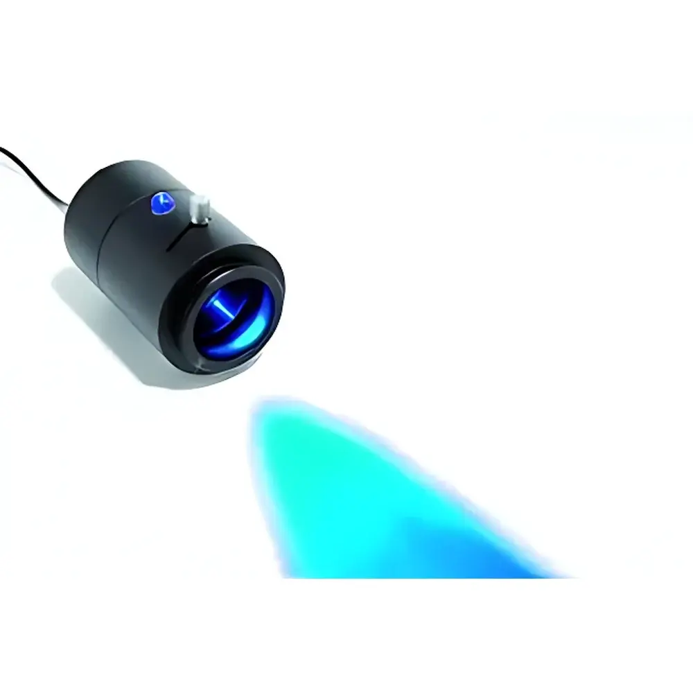
Leica SFL100 LED Fluorescence Excitation Light Source
| Brand | Leica |
|---|---|
| Origin | Germany |
| Model | SFL100 |
| Excitation Wavelength | 470 nm (standard), optional wavelengths available |
| LED Lifetime | ≥10,000 hours |
| Design | Compact, integrated, alignment-free illumination module |
| Power Control | Continuous, analog intensity adjustment |
| Compliance | CE, RoHS, IEC 61000-6-3 |
Overview
The Leica SFL100 is a compact, high-stability LED-based fluorescence excitation light source engineered for routine and research-grade fluorescence microscopy applications. Unlike traditional mercury or metal-halide arc lamps, the SFL100 employs solid-state 470 nm blue-light LEDs optimized for broad compatibility with common fluorophores—including FITC, GFP, Alexa Fluor 488, and SYTO dyes—while eliminating thermal drift, spectral instability, and hazardous UV emissions. Its core optical architecture is based on Köhler illumination principles, delivering uniform, collimated excitation light across the field of view without requiring manual lamp centering or warm-up time. Designed specifically for integration with Leica upright and inverted microscope platforms (e.g., DM series), the SFL100 operates as a drop-in replacement for conventional epifluorescence illuminators, preserving existing filter cube configurations while significantly improving operational reproducibility and user ergonomics.
Key Features
- Alignment-free LED illumination: No lamp centering, no focus calibration—enabling immediate use after power-on.
- Continuous analog intensity control: Precise, stepless dimming from 0–100% output without spectral shift or flicker, supporting quantitative intensity-dependent experiments.
- Thermally stabilized LED module: Active heat-sink design maintains consistent radiometric output over extended imaging sessions (>4 hours), minimizing photobleaching artifacts.
- 10,000-hour rated LED lifetime: Equivalent to >5 years of typical lab usage (8 h/day, 5 days/week), reducing downtime and consumables cost by >95% versus mercury lamps.
- Low electromagnetic interference (EMI) design: Compliant with IEC 61000-6-3 for co-location with sensitive electrophysiology or live-cell imaging setups.
- Compact footprint (120 × 85 × 65 mm): Enables benchtop deployment or integration into automated slide scanners and digital pathology workstations.
Sample Compatibility & Compliance
The SFL100 is validated for use with standard fluorescence filter sets (e.g., Leica L5, N2.1, Y5) and supports both fixed and live biological specimens—including formalin-fixed paraffin-embedded (FFPE) tissue sections, cytospin preparations, bacterial smears, and cultured mammalian cells. Its narrow spectral bandwidth (FWHM < 20 nm at 470 nm) ensures minimal crosstalk in multi-label experiments. The device meets CE marking requirements under the EU Medical Device Regulation (MDR 2017/745) for Class I non-invasive diagnostic equipment and conforms to ISO 13485:2016 quality management standards for in vitro diagnostic (IVD) accessory devices. It is routinely deployed in GLP-compliant histopathology labs and CLIA-certified clinical laboratories where traceable illumination stability is required for semi-quantitative IHC scoring.
Software & Data Management
While the SFL100 operates as a standalone hardware unit with manual intensity control, it is fully compatible with Leica Application Suite (LAS X) v3.7+ via TTL-triggered external control interface (BNC input). Integration enables synchronized LED activation with camera exposure timing, Z-stack acquisition, and multi-channel sequential imaging protocols. All intensity settings are logged within LAS X metadata (DICOM-SR compliant), supporting audit trails required under FDA 21 CFR Part 11 for regulated environments. No proprietary drivers or firmware updates are required—the device appears as a calibrated light source node within the microscope’s peripheral enumeration tree.
Applications
- Routine immunohistochemistry (IHC) screening in surgical pathology labs, where rapid turnaround and consistent signal-to-noise ratios improve diagnostic confidence.
- Microbiology workflows involving fluorescent in situ hybridization (FISH) and viability staining (e.g., BacLight), benefiting from stable excitation during long-duration image capture.
- Academic cell biology studies requiring low-phototoxicity illumination for time-lapse imaging of GFP-tagged cytoskeletal dynamics.
- Quality control in biomanufacturing, where standardized fluorescence intensity thresholds are used to verify antibody conjugation efficiency in QC lot release testing.
- Digital pathology scanner illumination modules, where spatial uniformity and temporal stability directly impact AI training dataset fidelity.
FAQ
Does the SFL100 support multiple excitation wavelengths?
Yes—while the standard configuration features a 470 nm LED, Leica offers factory-installed options at 365 nm (UV), 420 nm (violet), 505 nm (cyan), and 545 nm (green) upon order specification. Custom wavelength combinations require optical redesign and are subject to minimum order quantities.
Is the SFL100 compatible with non-Leica microscopes?
Mechanical and electrical integration is optimized for Leica DM series stands; third-party adaptation requires custom mounting brackets and independent power regulation—Leica does not validate or support such configurations.
What maintenance is required beyond LED replacement?
None. The sealed LED module contains no consumables, filters, or moving parts. Annual verification of output irradiance (mW/cm² at specimen plane) using a NIST-traceable photodiode sensor is recommended for ISO 17025-accredited labs.
Can intensity be controlled programmatically via serial command?
No—the SFL100 lacks RS-232 or USB-CDC interfaces. Intensity is adjusted exclusively via front-panel potentiometer or analog voltage input (0–5 V) for external DAQ synchronization.


