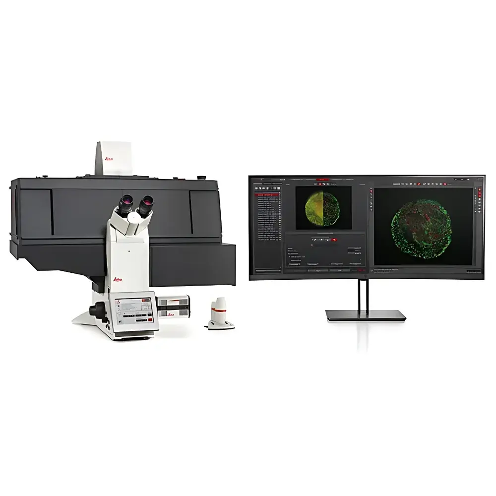
Leica THUNDER Imager 3D Live Cell Microscopy System
| Brand | Leica |
|---|---|
| Origin | Germany |
| Model | THUNDER Imager 3D Live Cell |
| Core Technology | Computational Clearing (real-time deconvolution for widefield fluorescence) |
| Imaging Modality | High-sensitivity widefield fluorescence microscopy with integrated environmental control |
| Camera | DFC9000 GTC sCMOS (QE up to 82%) |
| Illumination | Multi-line high-intensity LED source with narrow excitation bandwidths |
| Stage | Quantum motorized stage (≤ ±0.25 µm reproducibility |
| Focus Control | Adaptive Focus Control (AFC) + closed-loop Z-drive (20 nm repeatability) |
| Throughput | Up to 90 fps full-frame acquisition |
| Z-stack Speed | 150-layer stack in <60 s |
| Environmental Chamber | Integrated CO₂, temperature & humidity control with micro-humidifier for long-term live imaging |
| Software Platform | LAS X (GLP-compliant, audit trail enabled, FDA 21 CFR Part 11 ready) |
Overview
The Leica THUNDER Imager 3D Live Cell is an integrated widefield fluorescence microscopy platform engineered for quantitative, physiologically relevant 3D live-cell imaging. Unlike conventional confocal or spinning-disk systems, it leverages Leica’s proprietary Computational Clearing technology—a real-time, GPU-accelerated deconvolution algorithm that computationally removes out-of-focus blur directly during image acquisition. This enables high-fidelity optical sectioning without mechanical scanning or laser-based phototoxicity, preserving sample viability across extended time-lapse experiments. The system is built upon the fully automated DMi8 inverted microscope base, featuring a quantum motorized stage, closed-loop Z-drive, and environmental chamber certified for sustained CO₂-regulated, temperature- and humidity-stable culture conditions. Designed explicitly for advanced 3D models—including organoids, spheroids, retinal explants, and whole-tissue preparations—the THUNDER Imager delivers robust, reproducible data under true physiological constraints: minimal photon dose, sub-second temporal resolution, and nanometer-scale axial stability.
Key Features
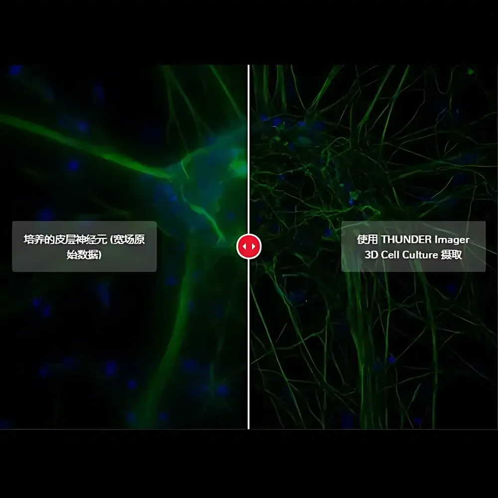
- Real-time Computational Clearing: Eliminates off-focus haze in widefield fluorescence images without iterative post-processing—enabling immediate visualization of intracellular detail at depth (up to 100 µm in cleared tissues).
- Ultra-low phototoxicity architecture: Narrow-band LED excitation sources synchronized with sCMOS shutter (20 µs switching) minimize cumulative light exposure; ideal for sensitive primary cultures and long-term developmental assays.
- High-throughput 3D acquisition: Full-frame sCMOS sensor (DFC9000 GTC) supports up to 90 fps acquisition; 150-plane Z-stacks completed in under 60 seconds with consistent signal-to-noise ratio across all layers.
- Physiological environment integration: On-stage incubation chamber maintains precise CO₂ (5%), temperature (±0.2°C), and relative humidity (>95%) over multi-day experiments; includes micro-humidifier for water-immersion objectives.
- Sub-micron positional fidelity: Quantum stage achieves ≤ ±0.25 µm positional reproducibility at speeds up to 10 positions/second; closed-loop Z-drive ensures ≤20 nm repeatability for longitudinal tracking of morphological changes.
- Adaptive Focus Control (AFC): Real-time hardware-based focus stabilization compensates for thermal drift, mechanical creep, and biological growth—critical for unattended multi-position time-lapse workflows.
Sample Compatibility & Compliance
The THUNDER Imager 3D Live Cell accommodates standard and custom 3D culture formats including glass-bottom dishes (e.g., #1.5 coverslip), Matrigel-embedded organoids, collagen- or fibrin-based hydrogels, and whole-mount tissue specimens (e.g., mouse retina, zebrafish pancreas). Its widefield illumination geometry and high-quantum-efficiency detection allow reliable imaging through plastic substrates—eliminating refractive index mismatches common in high-content screening plates. All hardware and software components comply with ISO 13485 design controls for medical device-related research instrumentation. LAS X software supports full audit trail logging, electronic signatures, and configurable user roles aligned with GLP and GMP documentation requirements. System validation packages include IQ/OQ documentation templates compliant with FDA 21 CFR Part 11 for regulated laboratories.
Software & Data Management
LAS X software serves as the unified interface for acquisition, analysis, and workflow automation. It provides native support for multi-channel, multi-position, multi-time-point experiments with programmable scheduling and conditional triggering. Built-in THUNDER processing modules apply Computational Clearing in real time or batch mode, with adjustable kernel size and regularization parameters optimized per sample thickness and fluorophore emission profile. Quantitative analysis tools include intensity-based segmentation, object counting (nuclei, organelles), colocalization (Pearson’s r, Mander’s coefficients), and 3D surface rendering with volume and surface area extraction. Raw data export conforms to OME-TIFF standards; metadata embedding follows MIAME and MINSEQE guidelines. Integration with third-party platforms (e.g., ImageJ/Fiji, Python via REST API, KNIME) enables scalable pipeline deployment in HPC or cloud-based analysis environments.
Applications
- Organoid development dynamics: Time-resolved tracking of neuroepithelial rosette formation in cerebral organoids using low-dose dual-color labeling; quantification of lumen expansion kinetics over 72 h.
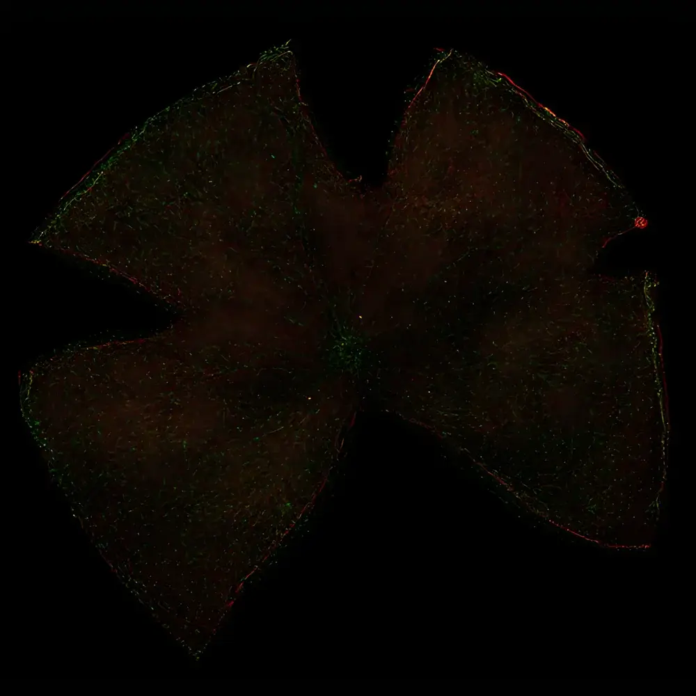
- Retinal phenotyping: Automated cell-type-specific enumeration (Iba1⁺ microglia, Brn3a⁺ RGCs) across full-thickness murine retinal sections with subcellular nuclear resolution.
- Metabolic trafficking studies: High-speed monitoring of insulin granule fusion events in INS-1 cells using TIRF-THUNDER hybrid mode—achieving 100 ms temporal resolution while maintaining widefield context.
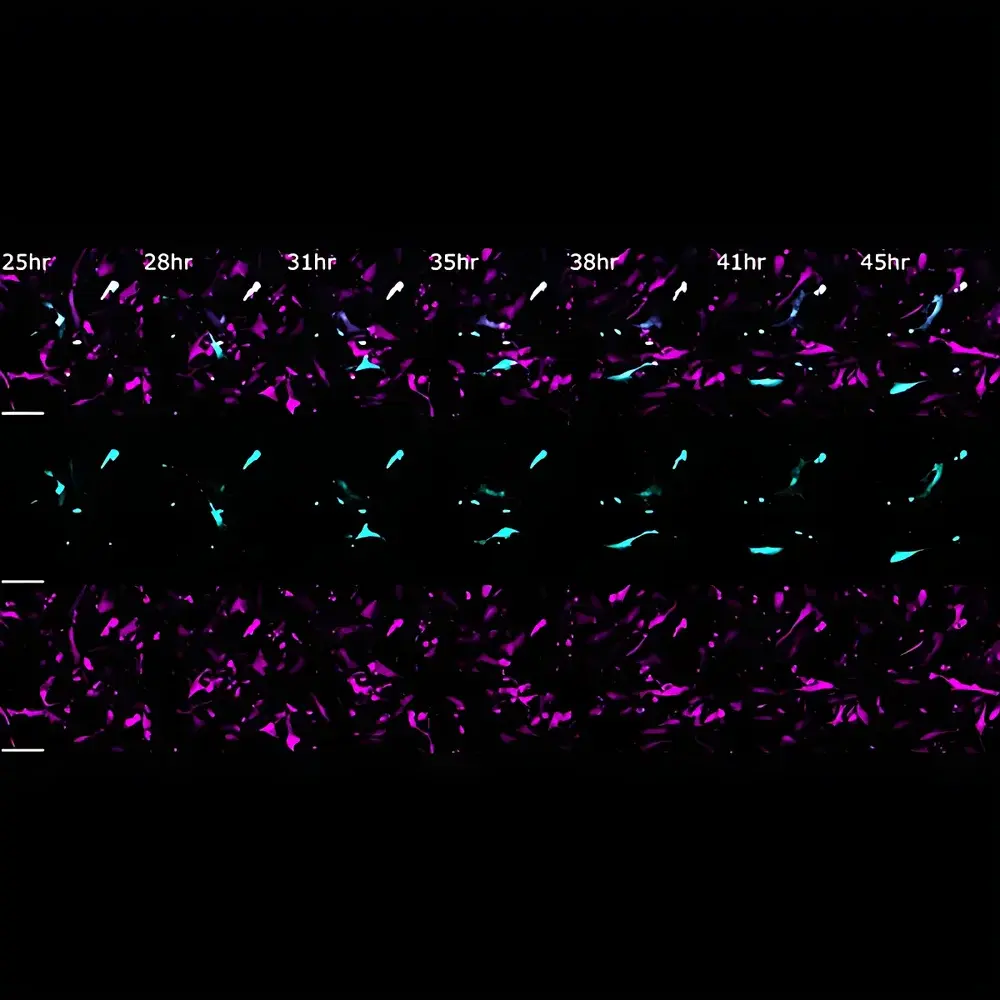
- Cardiovascular explant modeling: 48-hour uninterrupted imaging of aortic smooth muscle migration in gel-embedded explants, with adaptive focus correction compensating for tissue contraction.
- Chemical perturbation screening: Multi-well plate compatibility enables dose-response analysis of compound effects on spheroid growth, apoptosis, and vascular sprouting under physiological gas exchange.
FAQ
How does Computational Clearing differ from conventional deconvolution?
Computational Clearing operates in real time during acquisition using a pre-calibrated point spread function (PSF) model and GPU-accelerated constrained iterative restoration—avoiding the latency and memory overhead of post-hoc algorithms. It is optimized for live samples with dynamic refractive index variations.
Can THUNDER be used with standard tissue culture plastics?
Yes—the system’s optical path is corrected for common polystyrene and cyclic olefin copolymer (COC) substrates, enabling direct imaging without transfer to glass-bottom vessels.
Is LAS X software compatible with regulatory compliance requirements?
Yes—LAS X supports 21 CFR Part 11 compliance through electronic signature enforcement, immutable audit trails, role-based access control, and validated backup protocols.
What environmental parameters are actively controlled during imaging?
CO₂ concentration (0–20%, typical setpoint 5%), temperature (20–42°C, ±0.2°C stability), relative humidity (up to 98%), and optional O₂ modulation via external gas mixer integration.
Does THUNDER support correlative light-electron microscopy (CLEM) workflows?
Yes—OME-TIFF export preserves precise spatial metadata, and LAS X supports fiducial marker registration for seamless alignment with EM datasets acquired on compatible SEM/TEM platforms.


