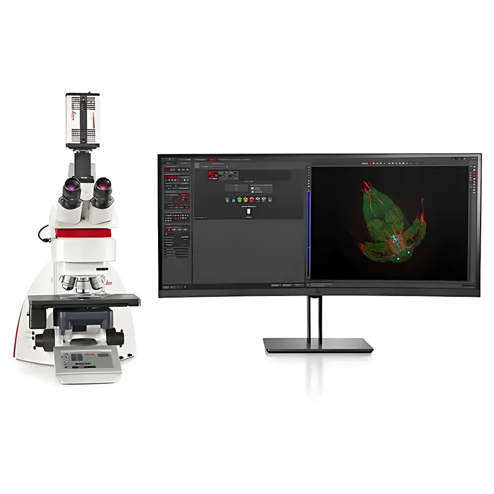
Leica THUNDER Imager Tissue Whole-Mount and Thick-Tissue Fluorescence Microscopy System
| Brand | Leica |
|---|---|
| Origin | Germany |
| Model | THUNDER Imager Tissue |
| Instrument Type | Upright/Inverted Hybrid Fluorescence Microscope |
| Excitation Source | High-Power LED Illumination |
| Classification | Research-Grade Widefield Fluorescence Microscope |
| Key Technology | Computational Clearing (Real-Time Deconvolution) |
| Optical Architecture | Optimized for 3D Tissue Imaging (up to 200 µm depth) |
| Software Platform | LAS X Navigator with Integrated THUNDER Engine |
| Compliance | Designed for GLP/GMP-aligned workflows |
Overview
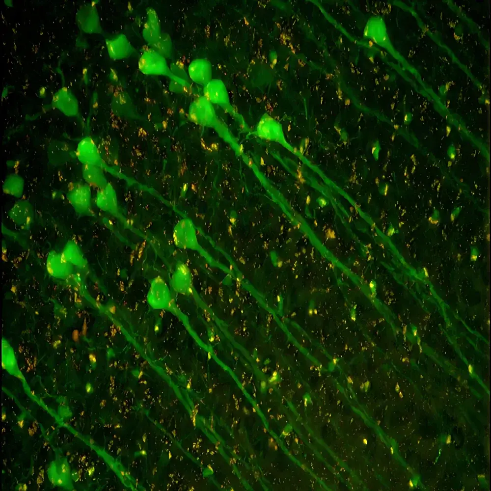
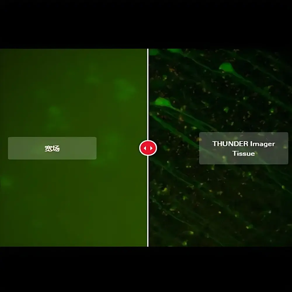
The Leica THUNDER Imager Tissue is a purpose-engineered widefield fluorescence microscopy system designed for high-fidelity, real-time imaging of intact biological tissues—including brain slices, organoids, whole-mount embryos, and cleared specimens—without the compromises traditionally associated with optical thickness. Unlike confocal or light-sheet systems that trade speed for sectioning depth, the THUNDER Imager Tissue leverages Leica’s proprietary Computational Clearing technology: a hardware-accelerated, pixel-level deconvolution algorithm applied in real time during acquisition. This approach eliminates out-of-focus blur while preserving native signal-to-noise ratio, photometric integrity, and acquisition speed—enabling true widefield advantages (e.g., high quantum efficiency, rapid field-of-view coverage, minimal phototoxicity) at depths previously accessible only via serial sectioning or point-scanning modalities. The system integrates a hybrid upright/inverted optical path, allowing flexible sample orientation—from delicate floating sections on glass slides to gravity-stable whole-mount preparations in specialized chambers—while maintaining consistent axial resolution and lateral uniformity across Z-stacks up to 200 µm thick.
Key Features
- Real-time Computational Clearing engine: On-the-fly deconvolution without post-processing delay or iterative parameter tuning; optimized for multi-channel fluorescence signals from common dyes (e.g., Alexa Fluor 488, Cy5, AF647) and fluorescent proteins (YFP, tdTomato)
- Dual-mode configuration flexibility: Choose between THUNDER Imager Tissue (manual focus, ideal for rapid overview imaging) or THUNDER Imager 3D Tissue (motorized Z-drive with sub-100 nm step precision, enabling reproducible volumetric acquisition)
- LED-based illumination architecture: Stable, cool, and spectrally tunable excitation sources with intensity modulation down to 0.1% increments; integrated neutral density filtering for quantitative intensity control
- Optimized optical train: High-transmission objectives (including 10×, 20×, and 40× water-dipping lenses with long working distances and corrected spherical aberration for thick samples)
- Automated fluorescence intensity management (FIM): Real-time adjustment of exposure, gain, and LED power to maintain linear response across dynamic range—critical for comparative quantification across large tissue areas
- Modular design compliant with ISO 9001-certified manufacturing standards; CE-marked and RoHS-compliant
Sample Compatibility & Compliance
The THUNDER Imager Tissue accommodates diverse specimen formats: free-floating vibratome sections (30–200 µm), paraffin-embedded or cryosections mounted on standard microscope slides, hydrogel-embedded tissues (e.g., CLARITY, iDISCO), and live ex vivo preparations maintained under physiological perfusion. Its open-stage architecture supports custom environmental chambers for temperature, CO₂, and humidity control. From a regulatory standpoint, the system meets essential requirements for research-use-only (RUO) instrumentation under IEC 61010-1 (safety) and IEC 62471 (photobiological safety). When paired with LAS X Navigator v4.13+ and the optional Security Pack, it supports ALCOA+ data integrity principles—including electronic signatures, role-based access control, and immutable audit trails—as required by GLP, GCP, and preclinical regulatory submissions aligned with FDA 21 CFR Part 11 and EMA Annex 11 guidelines.
Software & Data Management
Acquisition and analysis are unified within Leica’s LAS X platform, featuring native integration of the THUNDER Engine. LAS X Navigator enables tile-scan stitching of large tissue regions (>100 mm²) with sub-pixel registration accuracy and automatic focus map generation. All THUNDER-processed images retain full metadata—including objective ID, Z-step size, LED channel settings, and deconvolution kernel version—for FAIR (Findable, Accessible, Interoperable, Reusable) data stewardship. Export formats include OME-TIFF (with embedded XML metadata), HDF5 (for large-volume 3D datasets), and standardized N5 containers compatible with Bio-Formats and QuPath. Batch processing pipelines support automated segmentation of neuronal morphologies (e.g., Sholl analysis, branch point quantification) using user-defined or pre-trained deep learning models deployed via LAS X AI Extension.
Applications
This system is routinely deployed in neuroscience laboratories for mapping synaptic density in hippocampal or cortical layers, tracking axonal regeneration in spinal cord injury models, and visualizing microglial dynamics in Alzheimer’s disease tissue. In developmental biology, it enables rapid phenotypic screening of gene-edited embryo sections without sectioning artifacts. Histopathology cores use it for digital archiving of whole-slide immunofluorescence assays where multiplexed marker co-localization must be preserved across depth. Additionally, its speed and robustness make it suitable for core facility environments serving multiple users with heterogeneous expertise—requiring minimal training yet delivering publication-grade 3D reconstructions directly from acquisition.
FAQ
Does THUNDER Imager Tissue require a dedicated GPU or high-end workstation?
No. Computational Clearing runs on an embedded FPGA co-processor integrated into the camera interface board; standard Windows 10/11 workstations with ≥16 GB RAM and SSD storage meet minimum specifications.
Can THUNDER Imager Tissue be used with conventional immersion objectives?
Yes—provided they are Leica HC PL APO or HCX PL FL L series objectives with correction collars calibrated for the refractive index of your mounting medium (e.g., 1.38 for RIMS, 1.45 for FocusClear).
Is z-stack acquisition fully motorized in all configurations?
Only the THUNDER Imager 3D Tissue variant includes a precision piezo-driven Z-stage with closed-loop feedback; the base THUNDER Imager Tissue model uses manual coarse/fine focus controls.
How does Computational Clearing differ from classical deconvolution software?
Classical deconvolution requires iterative PSF estimation and offline computation, often introducing artifacts in low-SNR regions. THUNDER applies a deterministic, non-iterative algorithm trained on physically modeled PSFs and validated against gold-standard EM ground truth—ensuring consistency and reproducibility across users and sessions.
Can raw unprocessed images be saved alongside THUNDER-processed versions?
Yes. LAS X records both the original widefield frame and the THUNDER-cleared output in parallel, with synchronized timestamps and identical metadata tags for traceability.
Related Products


