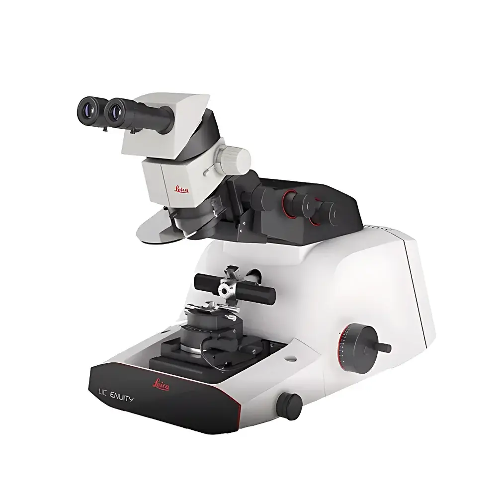
Leica UC Enuity Ultramicrotome
| Brand | Leica |
|---|---|
| Origin | Germany |
| Model | UC Enuity |
| Application | Electron Microscopy Sample Preparation |
| Temperature Modes | Room Temperature & Cryo-Sectioning |
| Integrated Fluorescence Monitoring | Yes |
| � | CT Data Integration: Yes |
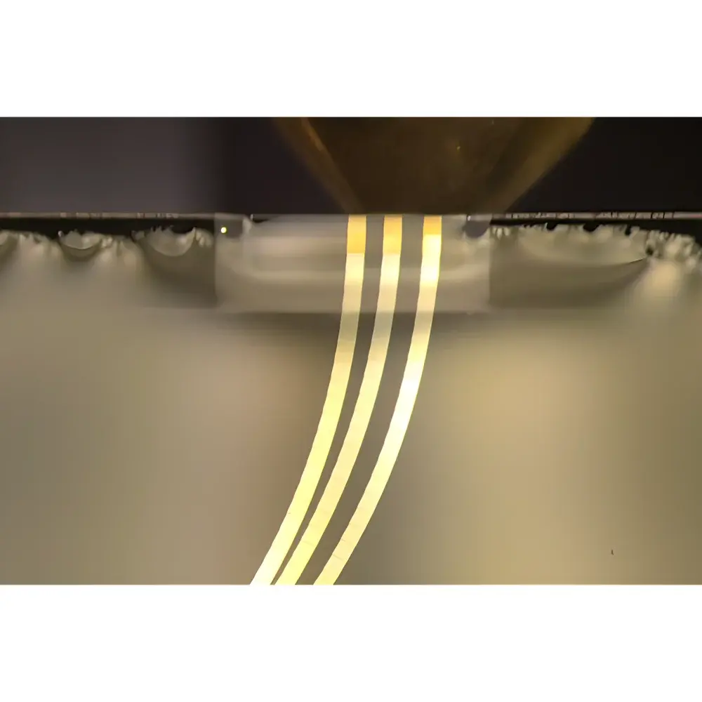
| Slice Thickness Range | 30 nm – 1 µm (typical for ultramicrotomy) |
| Motorized Block Advancement | Yes |
| Automated Trim Function | Yes |
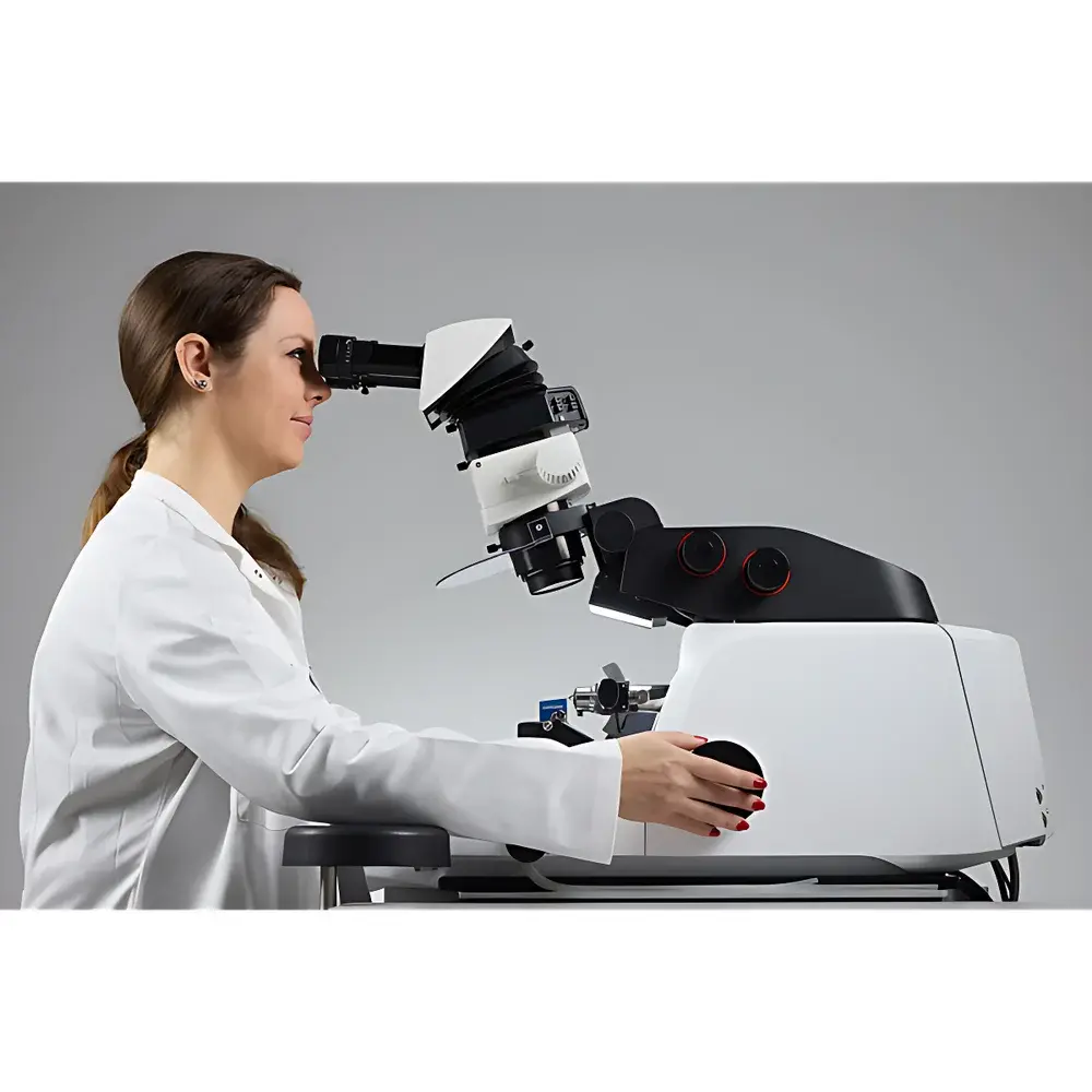
| Ergonomic Adjustable Stereo Microscope | Yes |
| Remote Diagnostics (RemoteCare) | Yes |
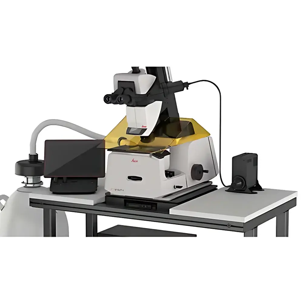
| Safety Features | UV-Blocking Shield, Closed LN₂ Handling System |
| Upgrade Path | Modular (Basic → Full Configuration) |
Overview
The Leica UC Enuity Ultramicrotome is an advanced, fully motorized instrument engineered for precision ultrathin sectioning in transmission electron microscopy (TEM) and serial block-face electron microscopy (SBEM) workflows. It operates on the principle of diamond-knife-based mechanical sectioning under controlled thermal and mechanical conditions, enabling reproducible generation of sections from 30 nm to 1 µm in thickness. Designed for both room-temperature resin-embedded sample sectioning and cryo-ultramicrotomy, the UC Enuity integrates real-time fluorescence monitoring, μCT data overlay, and automated block trimming—significantly reducing operator dependency while maintaining nanoscale geometric fidelity. Its architecture supports correlative light and electron microscopy (CLEM), array tomography, and high-throughput serial sectioning protocols essential for structural cell biology, neuroanatomy, and materials science applications.
Key Features
- Automated Block Trimming: Define region-of-interest boundaries via stereo microscope interface; system autonomously performs face trimming with sub-micron positional repeatability.
- In-situ Fluorescence Monitoring: Integrated LED-based epifluorescence illumination and filter set enables live fluorescence signal detection during both room-temperature and cryogenic sectioning—critical for CLEM-guided targeting.
- μCT Data Integration: Import and overlay micro-computed tomography datasets to spatially register internal structures prior to sectioning, facilitating precise navigation to deep-target regions without iterative trial cutting.
- Cryo-Optimized Thermal Management: Dual-zone temperature control (−160 °C to +40 °C) with integrated cryo-chamber and anti-icing airflow minimizes ice contamination during frozen-hydrated or freeze-substituted sample handling.
- Modular Expandability: Start with UC Enuity Basic configuration and incrementally add modules—including cryo-transfer station interface, electrostatic tape collector, silicon wafer auto-loader, and RemoteCare diagnostics suite—without hardware replacement.
- Ergonomic Human-Machine Interface: Fully articulating stereo microscope with height- and angle-adjustable armrests, padded forearm supports, and programmable focus presets reduce operator fatigue during extended sessions (>8 h).
Sample Compatibility & Compliance
The UC Enuity accommodates standard resin-embedded biological specimens (e.g., Epon, Araldite, Lowicryl), frozen-hydrated samples mounted on aluminum stubs, and semi-crystalline polymer blocks. It complies with ISO 13485:2016 for medical device manufacturing environments and supports GLP/GMP-aligned documentation through optional audit-trail logging (per FDA 21 CFR Part 11 when paired with Leica LAS X software). All optical components meet IEC 60825-1:2014 Class 1 laser safety standards; the UV-blocking shield satisfies EN 170:2002 requirements for ocular protection during fluorescence imaging.
Software & Data Management
Controlled via Leica Application Suite (LAS X) v4.13+, the UC Enuity provides synchronized acquisition of section metadata—including knife vibration amplitude, block advance step size, section count, ambient humidity, chamber temperature, and fluorescence intensity histograms. Raw data exports in TIFF/OME-TIFF format are compatible with Fiji/ImageJ, IMOD, and Amira for downstream 3D reconstruction. RemoteCare diagnostics continuously monitor over 30 real-time operational parameters and deliver predictive alerts for >20 fault codes—enabling proactive maintenance scheduling and minimizing unplanned downtime. Audit logs record user actions, parameter changes, and error events with timestamped digital signatures.
Applications
- Serial section TEM (ssTEM) for synaptic ultrastructure mapping in neuroscience
- Array tomography of immunolabeled tissue sections on silicon wafers
- Cryo-serial sectioning of vitrified cells for cellular tomography
- Correlative fluorescence-electron microscopy (CLEM) workflow integration
- Materials science analysis of composite thin films and battery electrode cross-sections
- Quality control of embedded semiconductor packaging interfaces
FAQ
What is the minimum achievable section thickness with the UC Enuity?
The instrument supports routine sectioning down to 30 nm using a diamond knife under optimized cryo or room-temperature conditions; thinner sections (≤20 nm) are feasible with specialized knife geometry and vibration-damping accessories.
Can the UC Enuity be integrated into an automated EM workflow?
Yes—it features TTL and Ethernet-based trigger I/O for synchronization with SEM/TEM stage controllers, autoloader systems, and downstream image acquisition platforms.
Is remote troubleshooting supported out-of-the-box?
RemoteCare functionality is embedded in firmware and requires only a secure LAN/WAN connection and Leica-certified remote access credentials.
Does the system comply with laboratory biosafety level (BSL) containment requirements?
While the UC Enuity itself is not classified as BSL-rated equipment, its closed cryo-chamber design and LN₂ containment pathway meet BSL-2 physical containment best practices for handling fixed biological specimens.
How is calibration traceability maintained?
Motorized advancement calibration is verified annually using NIST-traceable step gauges; fluorescence channel intensity is validated using Leica’s certified reference slide sets (Cat. No. 11507921).