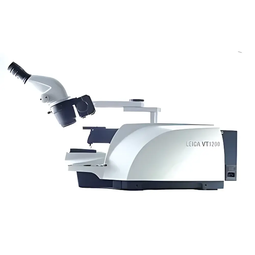
Leica VT1200 Semi-Automatic Vibratome
| Origin | Germany |
|---|---|
| Manufacturer Type | Authorized Distributor |
| Origin Category | Imported |
| Model | VT1200 |
| Pricing | Available Upon Request |
Overview
The Leica VT1200 semi-automatic vibratome is a precision-engineered instrument designed for the preparation of high-integrity, viable tissue sections from fresh, unfixed, or lightly fixed biological specimens—particularly in neuroscience research contexts. It operates on the principle of controlled vibrational cutting: a high-frequency oscillating blade moves vertically at adjustable amplitude and frequency while advancing incrementally through the specimen mounted on a magnetic stage. This mechanism minimizes shear stress and thermal damage, preserving cellular viability, membrane integrity, and electrophysiological functionality—critical for applications such as patch-clamp recordings, live-cell imaging, and acute slice electrophysiology. Developed in close collaboration with Prof. Dr. Peter Jonas’ laboratory at the Institute of Physiology, University of Freiburg, the VT1200 integrates neurobiological workflow requirements with mechanical stability and user-controlled reproducibility.
Key Features
- Semi-Automatic Operation Mode: Enables manual selection of section thickness (1–1000 µm, typical range 50–300 µm) and individual advancement per cut, granting full experimental control without reliance on pre-programmed protocols.
- Longitudinal Deviation Compensation: Equipped with the optional Vibrocheck detection system, the VT1200 allows real-time monitoring of blade trajectory deviation along the Z-axis; fine-tuning via micrometer-adjustable screws on the knife holder reduces longitudinal deviation to <1 µm—ensuring planar section uniformity and minimizing compression artifacts.
- Magnetic Sample Stage: Features a rare-earth magnet-based specimen holder that ensures rapid, repeatable, and secure positioning of agarose-embedded or gelatin-stabilized tissue blocks—reducing setup time and improving inter-slice alignment consistency across experiments.
- Vibration-Optimized Blade Drive: Utilizes a low-noise electromagnetic actuator delivering stable, sinusoidal oscillation (frequency range: 40–120 Hz) with amplitude adjustment (10–100 µm), enabling optimal cutting performance across soft neural tissues, embryonic samples, and fibrous peripheral tissues.
- Ergonomic & Modular Design: Includes adjustable specimen bath depth (up to 60 mm), removable stainless-steel chamber with integrated temperature control ports (compatible with recirculating chillers), and tool-free blade exchange mechanism compliant with standard vibrating microtome blades (e.g., Leica VT blades, Dosaka EM-type).
Sample Compatibility & Compliance
The VT1200 accommodates a broad spectrum of biological specimens—including rodent and human brain slices, spinal cord segments, retinal explants, hippocampal organotypic cultures, and developing embryonic tissues—without requiring cryofixation or paraffin embedding. Its non-contact, low-force cutting methodology maintains endogenous enzyme activity, receptor conformation, and synaptic ultrastructure, supporting downstream applications in immunohistochemistry, calcium imaging, and multi-electrode array (MEA) recording. The system complies with ISO 13485 design control principles for laboratory instrumentation and supports GLP-compliant documentation when integrated with validated electronic lab notebook (ELN) systems. While not a medical device, its operational parameters align with ASTM E2917-21 guidelines for vibratome performance verification in life science laboratories.
Software & Data Management
The VT1200 operates without proprietary software—its controls are fully hardware-based via front-panel dials and tactile switches—ensuring deterministic response, zero latency, and immunity to OS-level interruptions. All parameter settings (vibration frequency, amplitude, advance speed, section thickness) are mechanically indexed and physically recorded in lab notebooks per GLP practice. For auditability, users may integrate external digital loggers or time-synchronized video capture systems (e.g., via HDMI output from optional camera mounts) to document slicing events. No data storage, network connectivity, or FDA 21 CFR Part 11 electronic signature capability is included—consistent with its role as a stand-alone sample preparation tool rather than a regulated analytical platform.
Applications
- Acute brain slice preparation for in vitro patch-clamp and field potential recordings
- Live-tissue functional imaging (e.g., two-photon Ca2+ dynamics in dendritic spines)
- Preparation of thick sections (100–500 µm) for CLARITY, iDISCO, and other tissue-clearing protocols
- Electrophysiological studies requiring preserved synaptic transmission and intrinsic excitability
- Developmental neurobiology: slicing embryonic or postnatal CNS tissue with minimal mechanical disruption
- Pharmacological testing on native tissue architecture without fixation-induced epitope masking
FAQ
What types of tissue can be sectioned on the VT1200?
Fresh, unfixed, or lightly fixed (e.g., 4% PFA ≤ 2 h) neural, muscular, or epithelial tissues embedded in low-melting-point agarose (2–4%) or gelatin (5–7%). Cryopreserved or decalcified samples are not recommended.
Is the VT1200 compatible with chilled or heated slicing baths?
Yes—the specimen chamber accepts standard external temperature control units via inlet/outlet ports; operating temperature range: 0–40 °C, with typical use at 2–8 °C for electrophysiology.
Can I use third-party vibrating blades?
The VT1200 accepts standard 35-mm-wide vibrating microtome blades with 10-mm mounting height; compatibility confirmed with Leica, Dosaka EM, and Ted Pella blade formats.
Does the VT1200 require routine calibration?
No formal calibration is required; however, periodic verification of longitudinal deviation using Vibrocheck and mechanical zeroing of the advance mechanism are recommended per institutional SOPs.
Is service and technical support available outside Germany?
Yes—authorized Leica Microsystems service partners provide installation qualification (IQ), preventive maintenance, and on-site troubleshooting in over 60 countries, with documented service history traceable to serial number.


