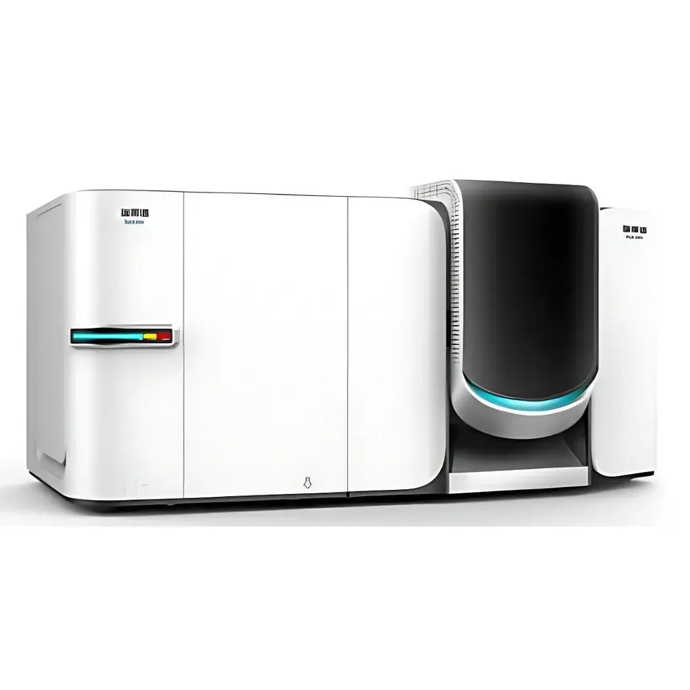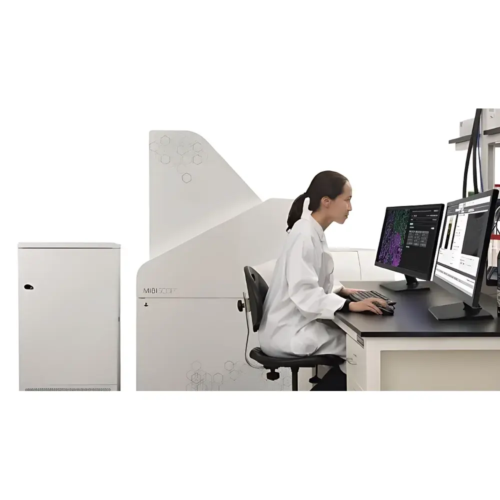
MIBIscope Spatial Proteomics Imaging System
| Origin | USA |
|---|---|
| Manufacturer Type | Distributor |
| Origin Category | Imported |
| Model | MIBIscope |
| Price Range | USD $720,000 – $1,200,000 |
| Instrument Type | Time-of-Flight (TOF) Mass Spectrometer |
| Application Scope | General-Purpose Biomolecular Imaging |
| Spatial Resolution | 350 nm – 1 µm |
| Sensitivity Ratio | >10:1 |
| Stability | Excellent |
| Data Acquisition Rate | 50 Gbit/s |
| Mass Accuracy | <80 ppm |
| Acquisition Time (Low Res, 1 µm) | 9–35 min |
| Acquisition Time (High Res, 500 nm) | 17–68 min |
| Acquisition Time (Ultra-High Res, 350 nm) | 35–139 min |
| Multiplex Channel Capacity | ≥40 Protein Markers |
| ROI Size | 400 × 400 – 800 × 800 µm² |
| Antibody Detection Limit | 1 ion (¹¹³In) to 16 ions (¹⁶⁶Er) |
| Dynamic Range | 5 decades (10⁵) |
| Output Format | TIFF |
Overview
The MIBIscope Spatial Proteomics Imaging System is a high-performance time-of-flight (TOF) mass spectrometry platform engineered for label-based, multiplexed ion beam imaging of formalin-fixed paraffin-embedded (FFPE) and frozen tissue sections. Leveraging Multiplexed Ion Beam Imaging (MIBI) technology, the system combines secondary ion mass spectrometry (SIMS) with metal-conjugated antibodies to deliver quantitative, subcellular-resolution protein expression maps across intact tissue architecture. Unlike conventional immunofluorescence or IHC methods limited by spectral overlap, MIBIscope enables simultaneous detection of ≥40 protein targets in a single acquisition—without signal crosstalk—by assigning each biomarker a unique stable isotope tag (e.g., ¹¹³In, ¹⁴²Nd, ¹⁶⁶Er). Its TOF mass analyzer provides high mass resolving power and <80 ppm mass accuracy, ensuring unambiguous elemental identification and robust discrimination between isotopic species. Designed for integration into routine pathology workflows, the MIBIscope operates under vacuum-compatible conditions and delivers spatially registered optical + secondary electron detector (SED) images for precise region-of-interest (ROI) definition prior to ion beam rastering.
Key Features
- True multiplex capability: Simultaneous quantification of ≥40 protein markers per tissue section using metal-tagged antibodies and isotopically resolved detection.
- Submicron spatial resolution: Adjustable pixel size from 350 nm to 1 µm, enabling visualization of cellular compartments and tumor microenvironment heterogeneity.
- High-fidelity mass detection: TOF mass spectrometer with <80 ppm mass accuracy and 5-decade dynamic range (10⁵), supporting absolute quantitation via internal calibration standards.
- Integrated multimodal imaging: Co-registered brightfield, fluorescence, and SED imaging for intuitive ROI selection and morphological context alignment.
- High-throughput data acquisition: Real-time streaming at 50 Gbit/s, enabling rapid generation of large-scale, multi-ROI TIFF datasets suitable for downstream computational pathology analysis.
- Workflow compatibility: Designed for use with standard FFPE tissue slides; minimal sample preparation beyond antibody staining and conductive carbon coating.
Sample Compatibility & Compliance
MIBIscope accepts standard 1–4 µm thick tissue sections mounted on conductive glass or silicon wafers. Compatible sample types include FFPE blocks, cryosections, and cytology smears. The system supports CLIA- and CAP-aligned laboratory practices and generates audit-ready metadata compliant with ISO/IEC 17025 requirements for analytical instrument validation. All acquired TIFF files retain embedded acquisition parameters (pixel size, dwell time, ion counts, calibration references), satisfying traceability mandates under FDA 21 CFR Part 11 when deployed in GLP/GMP environments. Antibody conjugation protocols follow manufacturer-recommended chelator chemistry (e.g., DOTA, NOTA) and are validated against ISO 13485–certified reagent suppliers.
Software & Data Management
The MIBIscope Control Suite provides instrument control, real-time ion counting, and automated calibration using reference standards (e.g., ¹⁰³Rh, ¹⁴⁰Ce). Acquired data are stored as lossless 32-bit TIFF stacks with per-pixel ion intensity values for each detected isotope. The配套 analysis platform—MIBI Analyst—supports spectral deconvolution, cell segmentation (via nuclear marker-guided clustering), phenotypic profiling, and spatial statistics (e.g., nearest-neighbor distance, neighborhood enrichment). Export formats include HDF5, CSV, and OME-TIFF for interoperability with QuPath, CellProfiler, and Python-based spatial transcriptomics pipelines. Audit logs record user actions, parameter changes, and calibration events—fully compliant with ALCOA+ principles for regulated research.
Applications
- Tumor microenvironment mapping: Characterization of immune cell infiltration patterns, stromal interactions, and checkpoint protein co-expression in oncology biopsies.
- Translational biomarker discovery: Validation of candidate protein signatures across large retrospective tissue cohorts with digital spatial context.
- Neurodegenerative disease profiling: Multiplexed detection of phosphorylated tau isoforms, amyloid-beta species, and glial activation markers in postmortem brain sections.
- Autoimmune tissue pathology: Spatial quantification of B-cell/T-cell colocalization, germinal center architecture, and cytokine receptor gradients in rheumatoid synovium or lupus nephritis specimens.
- Preclinical toxicology: Assessment of target engagement, off-target binding, and adaptive response markers in rodent and NHP tissue models.
FAQ
What sample preparation is required prior to MIBIscope analysis?
Standard immunostaining with metal-conjugated antibodies, followed by conductive carbon coating (10–20 nm thickness) to prevent charging during ion beam irradiation.
Can MIBIscope data be integrated with RNA-seq or spatial transcriptomics datasets?
Yes—TIFF-based ion intensity matrices are directly importable into Seurat, Squidpy, and SpatialDE for multimodal integration; coordinate-matched registration with Visium or Xenium data is supported via affine transformation tools.
Is mass calibration performed automatically during acquisition?
Yes—the system performs real-time mass calibration using internal reference ions (e.g., ¹⁰³Rh) every 100 µm of scan progression to maintain long-run stability and inter-session comparability.
How is data storage managed for large-scale studies involving hundreds of tissue sections?
The MIBIscope Data Server architecture supports tiered storage (SSD cache + NAS archive) and includes built-in DICOM-SR export for PACS integration in clinical research settings.
Does the system support Good Manufacturing Practice (GMP) documentation packages?
Yes—IQ/OQ/PQ protocols, 21 CFR Part 11-compliant electronic signatures, and full traceability reports are available through certified service agreements.