Micronit OOC Membrane Microfluidic Organ-on-a-Chip Chip
| Brand | Micronit |
|---|---|
| Origin | Netherlands |
| Model | ooc_membrane 00738, ooc_membrane 01206, ooc_membrane 01060 |
| Substrate Material | Borosilicate Glass |
| Membrane Material | PET |
| Membrane Thickness | 12 µm, 9 µm, 16 µm |
| Pore Size | 0.45 µm, 3 µm, 8 µm |
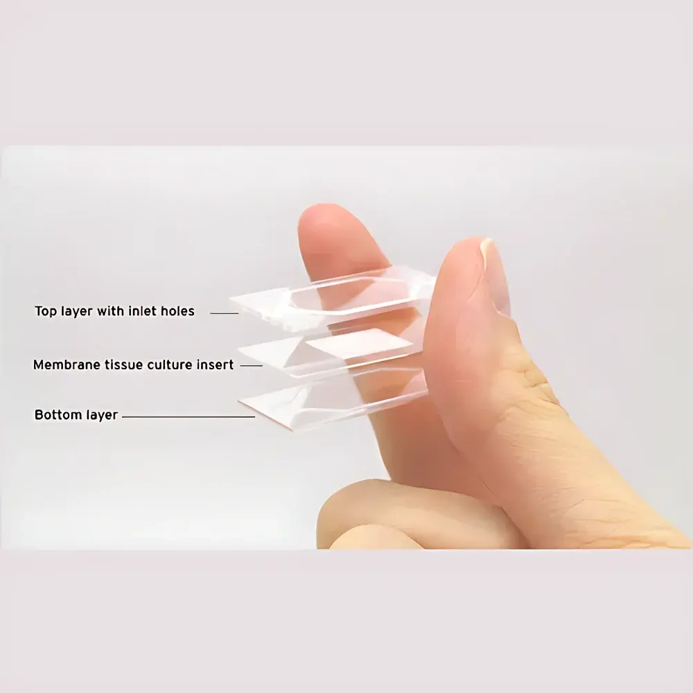
| Chip Architecture | Three-layer (glass baseplate, PET membrane, glass coverplate) |
| Sealing Method | Mechanical clamping with silicone gaskets |
| Compliance | Designed for ISO 13485-aligned cleanroom fabrication and GLP-compatible experimental workflows |
Overview
The Micronit OOC Membrane Microfluidic Organ-on-a-Chip Chip is a precision-engineered, reusable platform for physiologically relevant in vitro organ modeling. Based on a robust three-layer architecture—two borosilicate glass plates (base and cover) sandwiching a thin, microporous polyethylene terephthalate (PET) membrane—the chip enables spatially segregated dual-chamber culture under controlled fluidic conditions. This design replicates critical structural and functional features of native tissue interfaces, such as the epidermal-dermal barrier in skin models or the alveolar-capillary interface in lung models. The chip operates via passive or active microfluidic perfusion, supporting long-term (≥14-day) co-culture of primary cells, iPSC-derived lineages, or organoids while maintaining cell polarity, extracellular matrix deposition, and barrier integrity. Its modular geometry and standardized footprint ensure compatibility with commercial perfusion pumps, inverted microscopy, and high-content imaging systems.
Key Features
- Three-Layer Modular Design: Borosilicate glass baseplate and coverplate provide optical clarity (transmission >90% from 350–2000 nm), thermal stability (up to 120°C), and chemical resistance to ethanol, isopropanol, and aqueous buffers—enabling sterilization, reuse, and post-experiment immunostaining.
- Engineered PET Membranes: Available in three standardized configurations (ooc_membrane 00738, 01206, 01060) with precisely defined pore sizes (0.45 µm, 3 µm, 8 µm) and thicknesses (9–16 µm), optimized for selective molecular transport, trans-epithelial/endothelial electrical resistance (TEER) measurement, and mechanical compliance matching native tissues.
- Tooling-Compatible Sealing: Integrated silicone gasket grooves on both glass plates allow reliable, leak-free assembly using Micronit’s proprietary clamping fixtures—achieving sealing pressures up to 100 kPa without deformation or delamination, validated per ASTM F2621-20 for microfluidic device integrity.
- Reusability & Post-Analysis Accessibility: Non-permanent mechanical sealing permits full disassembly after culture, enabling downstream processing—including membrane harvesting for RNA extraction, histological sectioning, or TEM sample preparation—without compromising structural fidelity.
- Manufacturing Traceability: Each chip batch is produced in an ISO 13485-certified cleanroom (Class 7) in Enschede, Netherlands, with full lot-level documentation including surface roughness (Ra < 0.8 nm), membrane porosity uniformity (<5% CV), and endotoxin levels (<0.03 EU/mL).
Sample Compatibility & Compliance
The Micronit OOC Membrane Chip supports a broad spectrum of human and rodent cell types, including primary keratinocytes/fibroblasts (skin), human pulmonary microvascular endothelial cells (lung), Caco-2/HT29-MTX (gut), and iPSC-derived cardiomyocytes (heart). It is compatible with standard extracellular matrices (collagen I, fibronectin, Matrigel®) and routinely used in serum-free, xeno-free, and chemically defined media formulations. All materials comply with USP Class VI biocompatibility requirements and are free of detectable leachables per ISO 10993-12. The chip architecture conforms to FDA-recommended design controls for organ-on-a-chip devices intended for non-clinical safety assessment, and its data output structure supports alignment with OECD TG 497 (test guideline for alternative methods) and EMA/CHMP/ICH/2017/2019 guidance on non-animal models in drug development.
Software & Data Management
While the chip itself is hardware-only, it integrates seamlessly with industry-standard microfluidic control platforms—including Elveflow OB1 Mk III pressure controllers, Fluigent Flow-EZ modules, and Dolomite Mitos P-Pump systems—via standardized 1/4″-28 UNF threaded ports. Experimental metadata (flow rate, duration, medium composition) can be logged and synchronized with time-lapse imaging using open-source tools such as MicroManager or commercial packages like MetaMorph. For regulatory submissions, raw image stacks, TEER logs, and cytokine assay outputs generated using this chip may be archived with audit trails compliant with 21 CFR Part 11 when paired with validated LIMS or ELN solutions (e.g., LabArchives, Benchling).
Applications
- Reconstructing stratified epithelial barriers for dermatotoxicology and transdermal drug permeation studies
- Modeling vascularized tumor microenvironments with perfused endothelial monolayers and stromal co-cultures
- Studying immune cell transmigration across inflamed endothelial layers under physiological shear stress (0.5–4 dyn/cm²)
- Long-term functional assessment of air-liquid interface (ALI) cultures in respiratory disease modeling
- High-content screening of nanomaterial biodistribution and cytotoxicity across tissue barriers
FAQ
Can these chips be sterilized and reused?
Yes—autoclaving (121°C, 20 min), 70% ethanol immersion, or UV-C irradiation (254 nm, 30 min) are validated sterilization methods. Reuse is limited to ≤3 cycles to maintain gasket elasticity and membrane integrity.
What perfusion flow rates are recommended for barrier function assays?
For TEER measurements in skin or gut models, laminar flow at 1–10 µL/min is typical; higher rates (20–50 µL/min) are used for shear-dependent endothelial activation studies.
Is custom membrane pore size or geometry available?
Micronit offers custom MEMS-fabricated membranes under NRE agreements; lead time is 12–16 weeks with minimum order quantities applying.
Do the chips support real-time TEER monitoring?
Yes—integrated gold-plated electrodes are available as an optional upgrade (model suffix “-TEER”) with calibrated impedance readouts compatible with EVOM2 or CellZscope systems.
Are there application notes or protocol libraries available?
Micronit provides peer-reviewed SOPs, video protocols, and validation datasets for common organ models via their Technical Resource Portal—accessible upon institutional registration.


