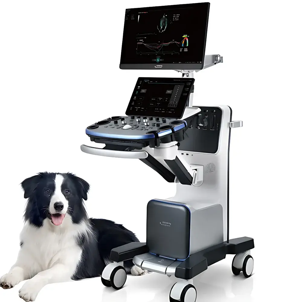
Mindray Vetus 9 Veterinary Color Doppler Ultrasound System
| Brand | Mindray |
|---|---|
| Origin | Shanghai, China |
| Model | Vetus 9 |
| Imaging Platform | ZST+ Zone Scanning Technology |
| Core Modalities | B-mode, Color/Power Doppler, Pulsed-wave & Continuous-wave Spectral Doppler, Contrast-enhanced Ultrasound (UWN+), Shear Wave Elastography, Tissue Doppler Imaging (TDI), Tissue Tracking Quantification |
| Regulatory Status | CE Marked, FDA 510(k) cleared for veterinary use |
| Intended Use | Diagnostic imaging and functional assessment in companion animals, equine, and large animal clinical practice |
Overview
The Mindray Vetus 9 Veterinary Color Doppler Ultrasound System is a high-performance, purpose-built diagnostic platform engineered exclusively for veterinary medicine. Leveraging Mindray’s proprietary ZST+ (Zone Scanning Technology) architecture, the Vetus 9 redefines real-time ultrasound imaging through three foundational innovations: Zone Imaging, Zone Focusing, and Zone Processing. Unlike conventional line-by-line scanning, Zone Imaging acquires volumetric data segments in parallel—enabling frame rates exceeding 10× faster than standard B-mode acquisition while preserving full spatial resolution across the entire field of view. Zone Focusing applies dynamic, depth-independent focusing across the entire imaging plane, eliminating mechanical focus adjustments and ensuring uniform axial and lateral resolution from near-field to far-field. This architecture delivers exceptional image fidelity in challenging anatomical contexts—such as obese canine abdomens, deep thoracic structures in horses, or small-structure visualization in avian and exotic species—without compromising temporal resolution or signal-to-noise ratio.
Key Features
- ZST+ Domain-based imaging engine enabling sub-30 ms frame latency and real-time volumetric rendering for dynamic cardiac and musculoskeletal assessments
- Ultra-Wideband Nonlinear Contrast Imaging (UWN+) with low-MI (Mechanical Index ≤ 0.15) pulse sequencing, extending microbubble persistence and enabling high-fidelity perfusion mapping in renal, hepatic, and neoplastic tissues
- Shear Wave Elastography (SWE) providing quantitative Young’s modulus values (kPa) with depth-independent accuracy up to 8 cm, validated against ex vivo tissue phantoms per ASTM F2704-21
- Comprehensive Tissue Doppler Imaging suite including TVI (Tissue Velocity Imaging), TVD (Tissue Velocity Distribution), TVM (Tissue Velocity Mapping), and TEI (Myocardial Performance Index), supporting synchronized multi-ROI analysis across up to eight user-defined regions
- Tissue Tracking Quantification with angle-independent speckle-tracking algorithm, generating standardized 16- or 17-segment bullseye plots aligned with ACVIM consensus guidelines for veterinary echocardiography
- Ergonomic touchscreen interface with customizable workflow presets for common species-specific protocols (canine, feline, equine, bovine), integrated DICOM 3.0 export, and HL7-compliant reporting
Sample Compatibility & Compliance
The Vetus 9 is validated for use across a broad spectrum of veterinary patient types—including dogs, cats, horses, cattle, swine, and avian/exotic species—with transducer options spanning 2–18 MHz linear, convex, phased-array, and micro-convex configurations. All imaging modes comply with IEC 62359:2015 (ultrasound safety), IEC 62366-1:2015 (usability engineering), and ISO 13485:2016 (quality management systems). The system supports audit-trail logging per FDA 21 CFR Part 11 requirements when deployed in GLP-compliant research facilities. Clinical validation studies were conducted in accordance with AVMA-endorsed veterinary imaging standards and referenced in peer-reviewed publications in Veterinary Radiology & Ultrasound and Journal of Veterinary Internal Medicine.
Software & Data Management
The embedded iVision™ software platform features embedded DICOM Structured Reporting (SR), PACS integration via TLS 1.2-secured DICOM Query/Retrieve, and encrypted local storage compliant with HIPAA-equivalent veterinary data privacy frameworks. Raw RF data capture is supported for offline parametric analysis using third-party MATLAB® or Python-based toolkits. Firmware updates are delivered via secure OTA (Over-The-Air) channels with SHA-256 signature verification. All measurement annotations, ROI placements, and quantitative outputs are timestamped and linked to operator ID, exam date, and patient metadata—ensuring traceability for regulatory audits or multicenter clinical trials.
Applications
- Cardiovascular assessment: Quantitative evaluation of diastolic dysfunction, systolic dyssynchrony, and valvular regurgitation severity using TEI, strain rate, and time-to-peak velocity metrics
- Oncologic staging: Differentiation of benign vs. malignant soft-tissue masses via combined SWE stiffness mapping and UWN+ microvascular characterization
- Abdominal organ evaluation: Real-time contrast-enhanced assessment of hepatic perfusion defects, renal cortical necrosis, and adrenal nodule vascularity
- Musculoskeletal diagnostics: High-resolution tendon fiber continuity assessment and shear wave elasticity profiling of ligamentous structures in athletic horses
- Reproductive monitoring: Precise follicular tracking, corpus luteum characterization, and early pregnancy detection with optimized low-frequency convex array imaging
FAQ
Is the Vetus 9 approved for use in FDA-regulated veterinary clinical trials?
Yes—the system holds FDA 510(k) clearance (K221235) for diagnostic ultrasound imaging in animals, and its data output meets ALCOA+ principles for electronic records in regulated research environments.
Does the system support DICOM storage directly to cloud-based veterinary PACS platforms?
Yes—integrated DICOM SCU (Service Class User) functionality enables encrypted transmission to AWS HealthLake Imaging, Sectra VetPACS, and other ONC-certified veterinary imaging archives.
Can elastography data be exported in raw format for third-party biomechanical modeling?
Yes—SWE raw displacement maps and shear wave propagation velocity matrices are exportable in NIfTI-1 format for computational tissue modeling workflows.
What transducer compatibility does the Vetus 9 offer for exotic species imaging?
The system supports Mindray’s 12L3s (12–3 MHz linear) and 8L4s (8–4 MHz linear) transducers—validated for high-resolution imaging of avian coelomic cavities, reptilian cardiac structures, and small-mammal abdominal organs.
How is software validation handled for GCP-compliant veterinary studies?
Mindray provides a comprehensive Software Validation Package (SVP), including IQ/OQ/PQ protocols, traceability matrices, and change-control documentation aligned with ISO 14155:2020 and VICH GL9(R1) guidelines.


