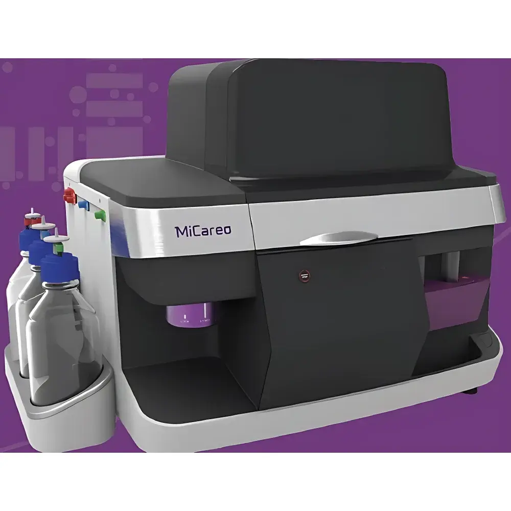
MiSelect R CTC Isolation and Analysis System
| Origin | Taiwan |
|---|---|
| Manufacturer Type | Distributor |
| Regional Category | Domestic (China) |
| Model | MiSelect R |
| Price Range | USD 280,000 – 420,000 |
| Type | Fully Automated |
| Sorting Speed | 100 cells/sec |
| Total Sortable Cells per Run | 10,000 |
| Sample Throughput | 0–6 samples/run |
| Sample Processing Time | 10–30 minutes |
| Programmable Protocols | 1 |
Overview
The MiSelect R CTC Isolation and Analysis System is a fully automated, integrated microfluidic platform engineered for the isolation, immunofluorescent labeling, high-resolution imaging, and recovery of viable circulating tumor cells (CTCs) directly from whole blood—without density gradient centrifugation, red blood cell lysis, or magnetic bead-based pre-enrichment. It operates on the proprietary eDAR (enhanced Droplet-based Antibody Recognition) technology: a nanoliter-scale droplet partitioning and real-time fluorescence-triggered sorting methodology. In eDAR, whole blood is precisely segmented into discrete nL-volume droplets; each droplet undergoes simultaneous surface marker labeling (including multiplexed antibody conjugates) within the microchannel. Upon optical interrogation via high-sensitivity excitation lasers, droplets exhibiting target-specific fluorescence signatures are electrostatically deflected onto a collection chip with sub-millisecond latency, while unlabeled droplets are directed to waste. This label-in-droplet, sort-at-source architecture preserves cell viability, minimizes shear stress, and eliminates manual handling-induced CTC loss—enabling downstream single-cell functional assays, RNA/DNA extraction, and NGS library preparation.
Key Features
- Fully automated end-to-end workflow—from raw whole blood input to image-verified, viable CTC recovery in under 30 minutes
- eDAR microfluidic engine enabling nanoliter-scale droplet generation, on-chip multiplex immunostaining, and fluorescence-activated droplet sorting (FADS) with 98.6% analytical sensitivity
- Real-time brightfield + dual-channel fluorescence imaging (488 nm & 561 nm excitation) for morphometric and phenotypic quantification of each isolated cell
- Integrated cell viability preservation: gentle hydrodynamic focusing, low-shear fluidics, and temperature-controlled processing chamber (maintained at 4–25 °C)
- Single-tube sample preparation: ≤5 minutes manual setup; no centrifugation, lysis, or antibody pre-incubation required
- Open-protocol compatibility: supports user-defined primary antibodies and fluorophore conjugates without system revalidation
Sample Compatibility & Compliance
The MiSelect R accepts native EDTA-anticoagulated whole blood (0.5–10 mL), bone marrow aspirates, and peripheral blood mononuclear cell (PBMC) suspensions. It complies with ISO 13485:2016 for in vitro diagnostic device design controls and supports audit-ready documentation per GLP and GCP frameworks. All image metadata—including timestamp, exposure parameters, droplet ID, fluorescence intensity histograms, and morphological descriptors—are embedded in DICOM-compliant output files. The system meets FDA 21 CFR Part 11 requirements for electronic records and signatures when deployed with validated LIMS integration.
Software & Data Management
Control and analysis are executed via the MiSelect Studio software suite (v3.2+), featuring a modular GUI with protocol builder, real-time event monitoring dashboard, and AI-assisted cell classification engine trained on >50,000 annotated CTC images. Raw image stacks (TIFF/OME-TIFF), droplet log files (.csv), and QC reports (PDF) are stored in a local encrypted SQLite database with optional SFTP or DICOM export. Audit trails record all user actions, parameter changes, and instrument status transitions with SHA-256 hashing. Software validation packages—including IQ/OQ/PQ documentation and traceability matrices—are provided for regulated environments.
Applications
- Early cancer detection and minimal residual disease (MRD) monitoring in liquid biopsy programs
- CTC enumeration and phenotypic profiling (e.g., EpCAM+/CK+/CD45−/DAPI+ with ≥13-plex co-expression analysis)
- Single-cell multi-omics preparation: whole-genome amplification (WGA), targeted RNA-seq, and methylation profiling
- Functional CTC characterization: live-cell migration assays, drug response testing, and co-culture with stromal cells
- Immunooncology research: concurrent isolation of CTCs and tumor-associated immune subsets (e.g., Tregs, MDSCs) from same sample
- Clinical trial biomarker qualification under CLIA-certified or CAP-accredited laboratory workflows
FAQ
What sample volume is required for optimal CTC recovery?
Minimum input is 0.5 mL whole blood; recommended range is 3–7 mL for robust statistical confidence in low-abundance CTC detection (≤1 CTC per 10⁷ WBCs).
Can the system isolate non-epithelial CTC subtypes (e.g., mesenchymal or stem-like)?
Yes—via user-defined antibody panels targeting vimentin, N-cadherin, CD44, or ALDH1A1; the eDAR platform does not rely on EpCAM capture and thus avoids epithelial bias.
Is recovered cell viability verified post-sort?
Yes—integrated trypan blue exclusion assay is performed automatically on collected fractions; typical post-sort viability exceeds 92% (n=42 clinical samples, mean ± SD).
Does the system support longitudinal tracking across multiple timepoints?
Yes—batch processing mode allows up to six samples per run with independent protocol assignment, enabling matched-pair analysis across serial draws.
How is data integrity ensured during regulatory submissions?
All acquisition parameters, image metadata, and sorting logs are digitally signed and archived with immutable timestamps; full chain-of-custody reporting is exportable in PDF/A-2b format compliant with EMA/ICH M10 guidelines.