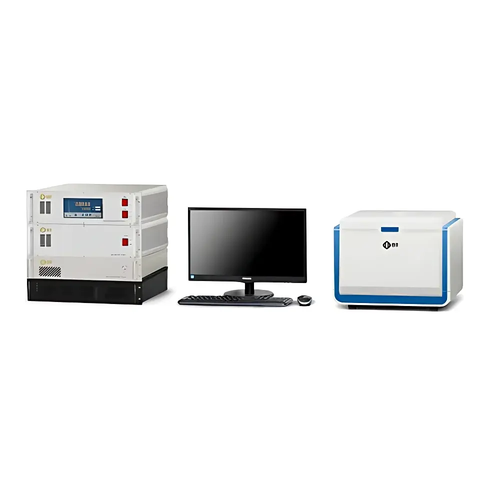
NIUMAG EDUMR-MRI Educational Benchtop Nuclear Magnetic Resonance Imaging System
| Brand | NIUMAG |
|---|---|
| Origin | Jiangsu, China |
| Model | EDUMR-MRI |
| Type | Benchtop NMR Imaging System for Education |
| Magnet Field Strength | 0.5 T (Permanent Magnet) |
| Operating Frequency | ~21.3 MHz (¹H) |
| Gradient Strength | ≥15 G/cm |
| RF Coil Options | Standard 25 mm & 50 mm Diameter Surface Coils |
| Data Interface | USB 3.0 + Ethernet |
| Software Platform | EDU-Imaging Suite v4.x with DICOM Export, K-Space Access, and Pulse Sequence Editor |
| Compliance | CE Marked, Meets IEC 61000-6-3/6-4 EMI/EMC Requirements |
| Safety Class | Class I, EN 61010-1 |
Overview
The NIUMAG EDUMR-MRI is a purpose-built, benchtop nuclear magnetic resonance (NMR) imaging system engineered exclusively for undergraduate and graduate education in medical physics, biomedical engineering, radiological sciences, and applied physics. Unlike clinical MRI scanners or research-grade NMR spectrometers, the EDUMR-MRI implements a permanent magnet-based 0.5 T platform operating at 21.3 MHz for proton (¹H) resonance — providing physically accurate signal generation, spatial encoding, and image reconstruction while maintaining classroom-safe footprint and operational simplicity. Its architecture follows the fundamental principles of Fourier-transform MRI: spin excitation via RF pulses, spatial localization using gradient fields, time-domain signal acquisition (FID and echo trains), and k-space-based image reconstruction. The system enables direct observation of Bloch equation dynamics — including T₁ relaxation, T₂ decay, spin echoes, and gradient refocusing — under controlled, repeatable experimental conditions. It is not intended for diagnostic use but serves as a pedagogically validated bridge between theoretical quantum mechanics and real-world MRI hardware operation.
Key Features
- Full hardware openness: All major subsystems — RF transmitter chain (pulse modulator, power amplifier), gradient driver circuits, receiver front-end (pre-amplifier, ADC), and spectrometer timing control — are accessible via test points and documented schematics for hands-on electronics labs.
- K-space data transparency: Raw, unprocessed k-space datasets are exported in MATLAB-compatible binary format and DICOM standard, enabling student-led reconstruction algorithm development (e.g., inverse FFT, SENSE, compressed sensing).
- Integrated virtual acquisition platform: EDU-Imaging Suite includes a real-time pulse sequence simulator that replicates scanner behavior — including gradient slew rate limits, RF pulse shape distortion, B₀ inhomogeneity, and thermal noise injection — allowing comparative analysis between simulated and acquired data.
- Multi-sequence capability: Native support for Spin Echo (SE), Fast Spin Echo (FSE), Inversion Recovery (IR), Gradient Echo (GRE), and Echo Planar Imaging (EPI) sequences — all configurable via intuitive GUI with adjustable TR, TE, TI, flip angle, bandwidth, and matrix size.
- Dual-mode operation: Simultaneous physical acquisition (using sample phantoms) and virtual acquisition (with parametric phantom models) enable scalable lab capacity — one instrument supports individual student workstations without hardware contention.
- Comprehensive pedagogical resources: Includes 22 structured lab modules aligned with ISO/IEC 17025-aligned assessment criteria, video-guided SOPs, instructor manuals with answer keys, and pre-lab quizzes embedded in LMS-compatible SCORM packages.
Sample Compatibility & Compliance
The EDUMR-MRI accommodates standard NMR teaching phantoms — including doped water, agarose gels, oil-water emulsions, and custom 3D-printed geometric inserts — with maximum sample diameter of 50 mm. It complies with IEC 61000-6-3 (emission) and IEC 61000-6-4 (immunity) for electromagnetic compatibility in educational environments. While not FDA-cleared or CE-certified as a medical device (per MDR 2017/745 Annex XVI), its design adheres to essential safety requirements outlined in EN 61010-1 for laboratory electrical equipment. All RF exposure levels remain below ICNIRP 2020 public exposure limits (≤ 0.08 W/kg whole-body SAR) during standard operation. Documentation includes full traceability of component-level conformity assessments and third-party EMC test reports.
Software & Data Management
EDU-Imaging Suite v4.x provides a unified interface for hardware control, real-time signal visualization (time-domain FID, frequency spectrum, echo train), k-space navigation, and image post-processing. The software supports audit-trail logging per GLP principles — recording user ID, timestamp, parameter set, and raw data hash for every acquisition. DICOM export (conformance statement available upon request) ensures interoperability with PACS training systems and third-party analysis tools (e.g., MITK, 3D Slicer). A dedicated Python API (PyEDUMR) enables integration with Jupyter notebooks for computational teaching — facilitating assignments on Fourier analysis, filter design, motion correction, and quantitative relaxometry. Data persistence follows ISO 15489-1:2016 guidelines, with optional encrypted local storage and network backup configuration.
Applications
The EDUMR-MRI supports curriculum delivery across multiple accreditation frameworks: ABET Criterion 3 (student outcomes), EFQM Higher Education Standards, and CAAHEP MRI Technologist competencies. In physics programs, it demonstrates quantum spin dynamics, resonance phenomena, and analog/digital signal processing. In biomedical engineering, students characterize SNR vs. voxel size, evaluate slice profile fidelity, and quantify geometric distortion from gradient nonlinearity. Medical imaging curricula utilize it for hands-on training in protocol optimization — e.g., how TE selection affects T₂-weighting, or how partial Fourier acquisition reduces scan time at cost of SNR. Research extensions include transverse relaxation mapping (T₂*), diffusion-sensitized sequence prototyping, and open-coil RF coil design validation. Optional add-ons — such as the RelaxoFit T₁/T₂ inversion recovery fitting module and 3D volume rendering toolkit — extend utility into capstone projects and faculty-led exploratory studies.
FAQ
Is the EDUMR-MRI suitable for clinical diagnostics?
No. It is strictly an educational instrument designed to replicate core MRI physics and engineering concepts — not certified for human use under FDA 21 CFR Part 820 or EU MDR.
Can students modify pulse sequence waveforms at the FPGA level?
Yes. The system includes documented VHDL source code for the digital spectrometer and a licensed Xilinx Vivado environment for advanced hardware-in-the-loop development.
Does the software support automated calibration routines?
Yes — including automatic RF center frequency search, gradient delay compensation, and quadrature detection phase alignment — all accessible via scriptable CLI mode.
What phantom materials are recommended for T₁/T₂ quantification labs?
NIUMAG-supplied MnCl₂-doped agarose gels (T₁: 200–2000 ms; T₂: 30–200 ms) and NiCl₂-doped water standards, with NIST-traceable reference values provided in the Lab Manual.
Is remote access supported for hybrid or distance learning?
Yes — via secure RDP over institutional VPN, with concurrent session throttling and role-based permissions (instructor/admin/student) enforced through integrated LDAP authentication.


