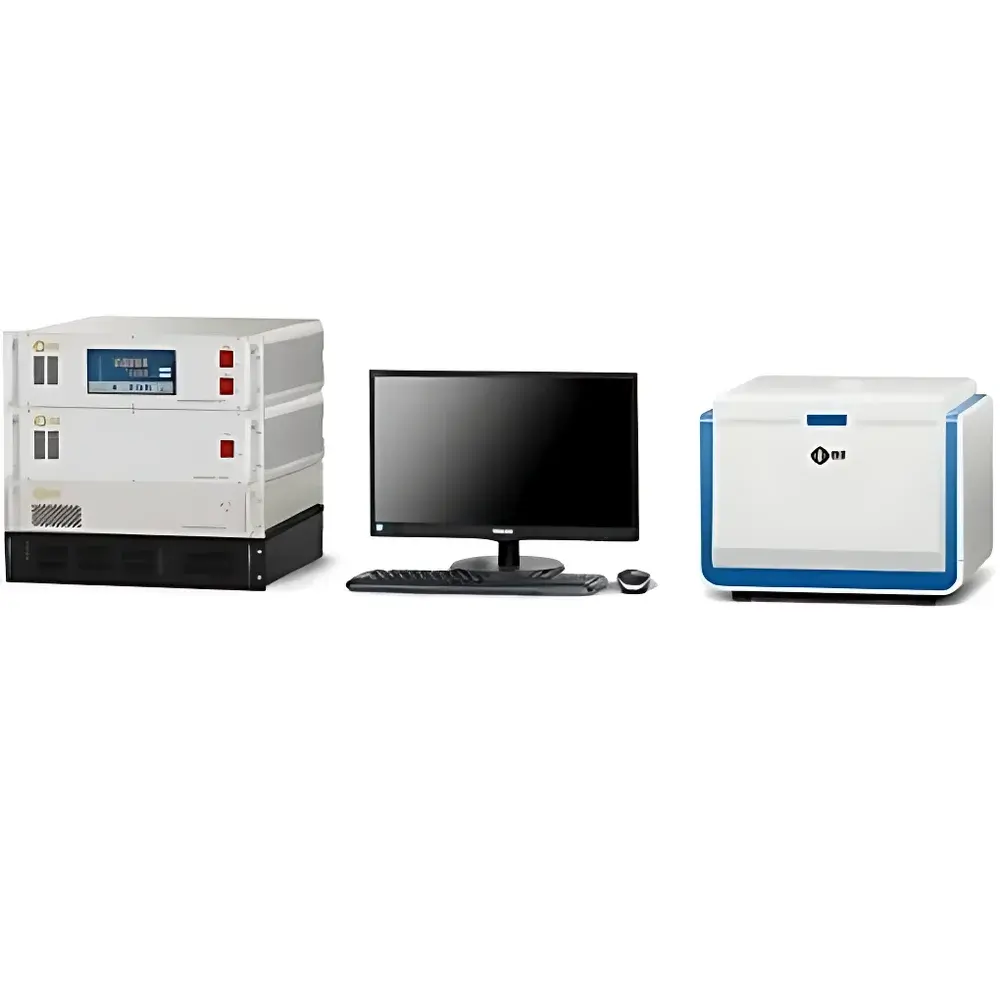
NIUMAG EDUMR20-015V Benchtop Low-Field Nuclear Magnetic Resonance Imaging Educational System
| Brand | NIUMAG |
|---|---|
| Origin | Jiangsu, China |
| Manufacturer Type | Authorized Distributor |
| Country of Origin | China |
| Model | EDUMR20-015V |
| Pricing | Available Upon Request |
Overview
The NIUMAG EDUMR20-015V is a compact, benchtop low-field nuclear magnetic resonance (NMR) imaging educational system engineered for pedagogical rigor and technical transparency in undergraduate and graduate physics, biomedical engineering, and medical imaging curricula. Unlike clinical MRI systems operating at 1.5–3 T, this instrument employs a permanent magnet with a nominal field strength of 0.5 T (21.3 MHz for 1H), enabling safe, accessible, and repeatable demonstration of core NMR principles—including spin excitation, RF pulse sequence timing, free induction decay (FID) acquisition, T1/T2 relaxation mapping, and k-space-based image reconstruction—without requiring shielded rooms or cryogenic infrastructure. Its architecture mirrors the functional modules of clinical MRI scanners (e.g., gradient coil assembly, RF transceiver, pulse programmer, and digital receiver), allowing students to map theoretical concepts directly onto physical hardware components.
Key Features
- Benchtop form factor with integrated permanent magnet (0.5 T), gradient coils (max. 0.15 T/m), and broadband RF probe (1H-tuned, 21.3 MHz)
- Fully open hardware interface: analog signal access points for oscilloscope/DAQ integration; real-time monitoring of RF pulses, gradient waveforms, and FID signals
- Open software architecture: raw k-space data export in MATLAB-compatible binary format (.dat); built-in pulse sequence editor supporting custom SE, GRE, and inversion-recovery sequences
- Dual-mode operation: continuous-wave (CW) NMR mode for foundational resonance experiments and pulsed NMR mode for imaging and relaxation studies
- Integrated teaching support: preloaded curriculum modules aligned with ISO/IEC 17025-compliant lab assessment frameworks, including safety protocols, calibration procedures, and uncertainty analysis guidelines
- Scalable deployment: supports multi-station classroom configurations via Ethernet-linked control nodes and synchronized data logging
Sample Compatibility & Compliance
The EDUMR20-015V accommodates cylindrical samples up to Ø25 mm × H50 mm, compatible with standard NMR tubes, tissue-mimicking phantoms (e.g., agarose gels doped with paramagnetic ions), and ex vivo biological specimens (e.g., rodent brain sections, plant stems). All RF and gradient subsystems comply with IEC 60601-2-33 (Safety Requirements for MRI Equipment) Class II limits for time-varying magnetic fields and SAR-equivalent exposure levels. The system meets GLP-aligned documentation standards, including electronic audit trails for sequence parameters, calibration logs, and user session records—facilitating compliance with academic accreditation requirements (e.g., ABET Criterion 3, IEEE Std 1012) and preparatory alignment with FDA 21 CFR Part 11 data integrity expectations.
Software & Data Management
Control and analysis are executed via NIUMAG’s EduMRI Studio v4.x—a cross-platform application built on Qt and Python (NumPy, SciPy, PyDicom). It provides real-time visualization of FID decay curves, k-space filling trajectories, and reconstructed magnitude/phase images (FFT-based or iterative SENSE reconstruction). Raw k-space datasets include metadata headers specifying TR/TE/TI, FOV, matrix size, and gradient amplitude profiles. Export formats include DICOM (conformance class MR Image Storage SOP), NIfTI-1, and HDF5—enabling interoperability with open-source tools such as FSL, AFNI, and MITK. Built-in scripting API supports automated batch processing, algorithm validation (e.g., compressed sensing reconstruction), and integration into Jupyter-based computational labs.
Applications
- Undergraduate physics labs: Larmor precession demonstration, resonance frequency tuning, and Bloch equation modeling
- Biomedical engineering courses: T1/T2 mapping of phantoms, diffusion-weighted imaging (DWI) simulations, and contrast agent quantification
- Radiologic technology training: slice selection gradient characterization, RF pulse calibration, and artifact analysis (e.g., chemical shift, magnetic susceptibility)
- Graduate research: development of novel reconstruction algorithms, motion correction strategies, and low-field-specific denoising techniques
- Cross-disciplinary projects: materials science (polymer gel swelling kinetics), food science (moisture distribution in grains), and plant physiology (xylem water transport imaging)
FAQ
Does the EDUMR20-015V support quantitative T1 and T2 mapping?
Yes—preconfigured inversion-recovery and multi-echo spin-echo sequences enable pixel-wise T1/T2 fitting using mono-exponential decay models, with uncertainty propagation calculated per ISO/IEC Guide 98-3.
Can students modify pulse sequences without vendor approval?
Yes—the embedded pulse programmer accepts user-defined C++-compiled sequence binaries; full source templates and timing constraint documentation are provided under academic license.
Is DICOM export compliant with PACS integration standards?
Yes—generated DICOM objects conform to PS3.3 (Information Object Definitions) and PS3.10 (Media Storage and File Format), including mandatory tags for modality (MR), manufacturer (NIUMAG), and institutional identification.
What safety certifications does the system hold?
It carries CE marking under Directive 2014/30/EU (EMC) and 2014/35/EU (LVD); magnetic field emission testing reports per EN 62233:2008 are available upon institutional request.
How is system calibration maintained across multiple student workstations?
Each unit includes an NIST-traceable reference phantom (CuSO4 solution); automated calibration routines verify RF center frequency, gradient linearity, and receiver gain stability before each experimental session.


