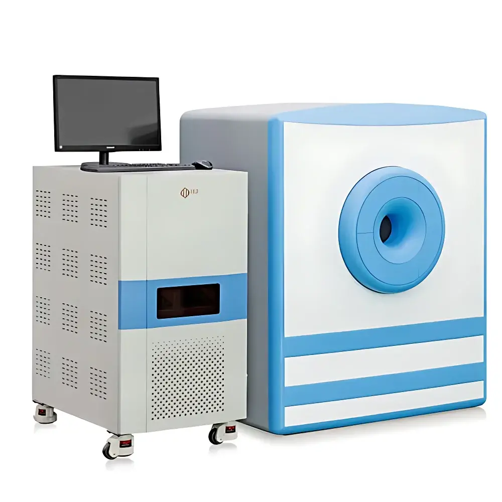
NIUMAG NM42-MRI Small Animal Magnetic Resonance Imaging System
| Brand | NIUMAG |
|---|---|
| Origin | Jiangsu, China |
| Manufacturer Type | Authorized Distributor |
| Country of Origin | China |
| Model | NM42-MRI |
| Pricing | Available Upon Request |
Overview
The NIUMAG NM42-MRI is a compact, high-performance benchtop magnetic resonance imaging system engineered for preclinical research in small animal models. Operating at a stable 1.0 Tesla permanent magnet field, the system leverages conventional spin-echo and gradient-echo pulse sequences to generate high-fidelity T1-, T2-, and proton density-weighted images without ionizing radiation. Its homogeneous static magnetic field (≤15 ppm over 40 mm DSV) ensures robust signal-to-noise ratio (SNR) and spatial resolution down to 80 µm in-plane with slice thickness adjustable from 0.8 mm upward—enabling precise anatomical delineation and longitudinal monitoring in mice, rats, and other rodents weighing between 1 g and 45 g. Designed for non-invasive, repeatable in vivo assessment, the NM42-MRI supports quantitative MRI biomarker extraction—including transverse relaxation time (T₂), longitudinal relaxation time (T₁), and apparent diffusion coefficient (ADC)—critical for evaluating therapeutic response, tumor burden progression, and contrast agent pharmacokinetics.
Key Features
- 1.0 T permanent magnet architecture with active shimming: Delivers consistent field homogeneity across a 40 mm spherical volume, enabling reproducible image acquisition and quantitative relaxometry.
- Non-ionizing, non-invasive operation: Eliminates radiation exposure risk and physiological stress associated with X-ray or PET modalities—ideal for longitudinal studies requiring repeated imaging sessions.
- Streamlined workflow: Fully automated parameter optimization; three-step imaging protocol (position → select sequence → acquire) reduces operator dependency and training time.
- Modular expandability: Optional integrated gas anesthesia delivery system (isoflurane-compatible) with real-time physiological monitoring interface (respiratory gating support).
- Low infrastructure footprint: No RF-shielded room required; operates under standard laboratory conditions (ambient temperature 15–30 °C, humidity <70% RH, non-condensing); minimal maintenance and zero consumables.
- Dual-mode capability: Supports both in vivo small animal imaging and high-throughput in vitro analysis of nanomaterial suspensions, ionic solutions, and microbial cultures (minimum sample volume: 100 µL).
Sample Compatibility & Compliance
The NM42-MRI accommodates specimens within a 40 mm transverse field-of-view (FOV), compatible with standard mouse and juvenile rat imaging cradles. In vitro samples must be contained in MR-compatible tubes (e.g., 10 mm OD glass or polypropylene) and exhibit sufficient proton density for detection. The system complies with IEC 61000-6-3 (EMC emission standards) and IEC 61000-6-2 (immunity). While not FDA-cleared for human diagnostics, it meets ISO 13485-aligned quality management practices for preclinical instrumentation. Data acquisition and storage support audit-trail functionality compatible with GLP-compliant workflows; optional software modules provide 21 CFR Part 11–ready electronic signatures and user access controls.
Software & Data Management
The NM42-MRI ships with two integrated software suites: the NIUMAG MRI Acquisition Suite and the NIUMAG Image Processing Toolkit. The Acquisition Suite provides full pulse sequence configurability—including TR/TE adjustment, flip angle tuning, echo train length, and k-space sampling schemes—while maintaining intuitive graphical controls. All parameters are saved with DICOM-compliant headers (including manufacturer, model, field strength, sequence type, and acquisition date). The Image Processing Toolkit enables ROI-based quantification (T₁/T₂ mapping, ADC calculation), multi-slice co-registration, pseudo-color rendering, 3D surface reconstruction, distance/angle measurement, threshold segmentation, and batch export to CSV, NIfTI, or MATLAB formats. Both applications support Windows 10/11 (64-bit) and integrate with third-party platforms such as ImageJ/Fiji via open API protocols.
Applications
- Contrast agent relaxivity characterization: Quantitative r₁ and r₂ determination in phantoms and in vivo murine models per ASTM E2925 and ISO 10993-18 guidelines.
- Oncology research: Longitudinal tumor volume tracking, necrosis assessment via T₂ mapping, and treatment efficacy evaluation following chemotherapy or immunotherapy regimens.
- Nanomedicine development: Rapid screening of nanoparticle colloidal stability, payload release kinetics, and biodistribution profiles using T₂*-weighted susceptibility contrast.
- Neuroscience: High-resolution structural imaging of brain anatomy, stroke lesion evolution, and white matter integrity assessment in transgenic models.
- Microbiology: Detection and quantification of bacterial load in biofilm or planktonic suspensions through proton density and diffusion-weighted signal attenuation.
FAQ
Does the NM42-MRI require a dedicated RF-shielded room?
No. Its permanent magnet design and optimized RF coil shielding enable operation in standard laboratory environments without additional Faraday cage construction.
Can the system perform diffusion-weighted imaging (DWI)?
Yes. The gradient subsystem supports b-values up to 2000 s/mm², enabling robust ADC map generation for tissue microstructure analysis.
Is anesthesia integration mandatory for in vivo imaging?
Not mandatory—but strongly recommended for motion artifact suppression during scans exceeding 2 minutes. The optional isoflurane delivery module includes vaporizer, flow meter, and scavenging interface.
What file formats are supported for data export?
DICOM (Level 3 conformance), NIfTI (.nii), Analyze (.hdr/.img), and CSV for ROI statistics; raw k-space data is accessible via MATLAB-compatible binary export.
How frequently does the magnet require recalibration?
The permanent magnet requires no routine recalibration. Field homogeneity is verified annually using built-in phantom-based QA routines compliant with ACR MRI Quality Control Manual recommendations.


