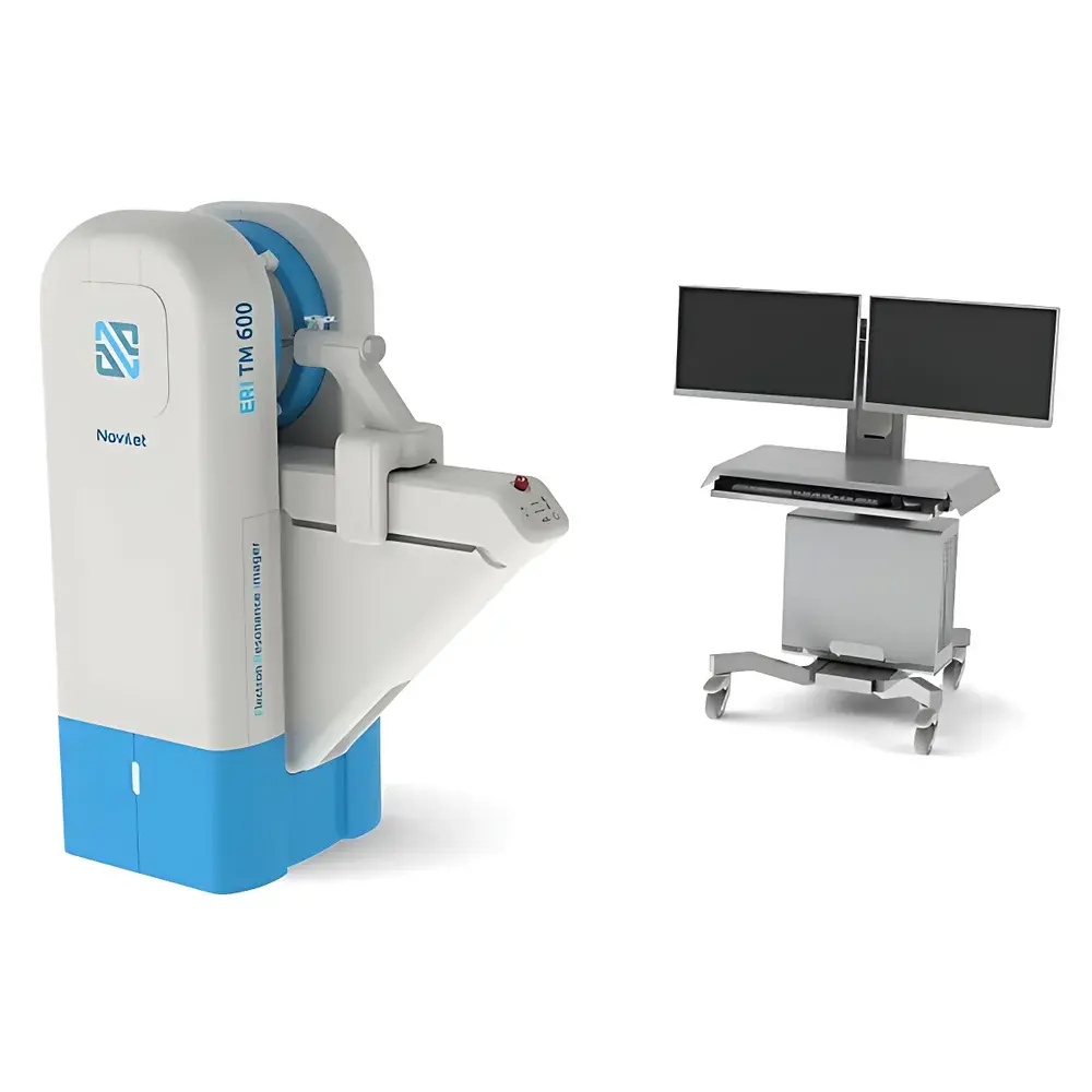
Novilet ERI TM 600 Small Animal In Vivo Electron Paramagnetic Resonance Imaging System
| Brand | Novilet |
|---|---|
| Origin | Poland |
| Model | ERI TM 600 |
| Imaging Modality | Electron Paramagnetic Resonance (EPR) Imaging |
| Sample Type | Live small animals (mouse, rat) |
| Spin Probe Required | Yes |
| Field Strength Range | 0.01–500 G (CW), 0.01–50 G (rapid scan) |
| Operating Frequency | 575 MHz |
| RF Power Output | Up to 500 mW |
| Modulation Amplitude | Up to 50 G |
| Modulation Frequency | 1 kHz |
| Magnetic Field Resolution | 10 mG |
| Field Stability | ±20 mG |
| Field Noise | ≤5 mG RMS |
| Gradient Strength | 13 G/cm |
| Magnet Bore Diameter | 34 mm |
| Imaging Volume | 20 cm³ |
| 3D Acquisition Time | As low as 4.5 s (225 projections) |
| SNR (OXO63, 25 g mouse, 100 ms) | 112 |
| Digital Resolution | 16-bit |
| Q-Factor | >800 |
| Temperature-Controlled Animal Holder | Integrated |
| Anesthesia-Compatible Gas Exchange System | Included |
| Stereotactic Mouse Brain Fixation | Standard |
Overview
The Novilet ERI TM 600 is a dedicated in vivo electron paramagnetic resonance (EPR) imaging system engineered for non-invasive, quantitative mapping of paramagnetic species—including endogenous and exogenous free radicals, oxygen partial pressure (pO₂), redox status, pH, and reactive oxygen species (ROS)—in live small animals. Unlike optical or nuclear imaging modalities, EPR imaging directly detects unpaired electrons, offering intrinsic specificity to paramagnetic centers without background signal from biological tissue. The system operates at 575 MHz using a high-Q resonator (Q > 800) and a precisely controlled permanent magnet with active field stabilization (±20 mG drift over 1 h), enabling high-fidelity spectral-spatial encoding. Its rapid-scan and multi-harmonic acquisition modes support dynamic 3D tomographic reconstruction with temporal resolution down to 4.5 seconds per volume—critical for tracking pharmacokinetics, metabolic fluxes, and real-time oxygen dynamics in tumor microenvironments or neurodegenerative models.
Key Features
- High-sensitivity in vivo EPR imaging optimized for murine and rat models, with integrated stereotactic brain fixation and temperature-regulated animal chamber (37 °C ± 0.3 °C)
- Dual-mode magnetic field control: continuous-wave (CW) for high-resolution pO₂ mapping (0.01–500 G range) and rapid-scan mode (0.01–50 G) for accelerated dynamic imaging
- Low-noise field generation (<5 mG RMS noise) and sub-10 mG field resolution ensure reproducible quantification of spin probe linewidth broadening—directly correlated to local pO₂
- Modular resonator design with 34 mm bore accommodates physiological instrumentation (IV lines, respiration monitoring, gas anesthesia delivery) during acquisition
- Real-time spectral acquisition (1 ms–10 s per spectrum) and on-board spectral deconvolution for multi-component spin probe analysis (e.g., OXO63, trityl, nitroxides)
- Automated 3D back-projection reconstruction engine supporting filtered back-projection (FBP), iterative SART, and Tikhonov-regularized algorithms
Sample Compatibility & Compliance
The ERI TM 600 is validated for use with standard preclinical spin probes including lithium phthalocyanine (LiPc), OXO63, and triarylmethyl (TAM) derivatives. It supports intravenous, intraperitoneal, and stereotactically guided intracerebral probe administration. The animal handling module complies with OECD Guidelines for the Testing of Chemicals (Section 4) and adheres to ISO/IEC 17025 principles for measurement traceability. All hardware timing, field calibration, and data logging modules are designed to meet GLP audit requirements, including full electronic record retention, user access controls, and audit trail functionality for raw spectral files, reconstruction parameters, and metadata (per FDA 21 CFR Part 11 Annex 11 expectations). No ionizing radiation is involved; operation requires only standard laboratory biosafety level 2 (BSL-2) infrastructure.
Software & Data Management
Acquisition, reconstruction, and quantification are managed via Novilet EPR Studio—a modular, Windows-based platform built on Qt and Python 3.9. Core modules include: (i) PulseSequence Designer for custom CW/rapid-scan waveform definition; (ii) SpectralFit Engine implementing nonlinear least-squares fitting of Lorentzian/Gaussian line shapes with automatic baseline correction; (iii) TomoRecon Suite supporting DICOM export (EPR-RT extension), NIfTI-1 compatibility, and co-registration with CT/MRI via landmark-based affine transformation. All datasets are stored in HDF5 format with embedded MIAME-compliant metadata (probe identity, dose, time post-injection, physiological parameters). Raw data integrity is ensured via SHA-256 checksums; version-controlled processing pipelines enable full retraceability from raw FID to quantitative pO₂ maps.
Applications
- Quantitative tumor hypoxia mapping: Direct pO₂ measurement in orthotopic and subcutaneous xenografts (e.g., LNCaP prostate cancer), enabling correlation with HIF-1α expression and radiotherapy response prediction
- Neurodegeneration redox profiling: Spatial mapping of ascorbyl radical and nitric oxide adducts in APP/PS1 and α-synuclein transgenic models to assess regional oxidative stress burden
- Ischemia-reperfusion injury kinetics: Real-time tracking of ROS burst magnitude and spatial spread in middle cerebral artery occlusion (MCAO) models
- Pharmacodynamic assessment of antioxidant therapeutics: Longitudinal monitoring of catalase-mimetic probe clearance rates and redox buffer capacity in liver and kidney
- Multimodal validation: Co-registered EPR/ultrasound and EPR/CT workflows for anatomical anchoring—demonstrated in skull surface localization studies and bone-adjacent tumor margin delineation
FAQ
Does the ERI TM 600 require radioactive tracers or ionizing radiation?
No. EPR imaging relies solely on exogenous or endogenous paramagnetic species and uses non-ionizing radiofrequency energy at 575 MHz.
Can the system image endogenous radicals without spin probes?
Yes—under specific pathological conditions (e.g., ischemic stroke, radiation exposure), endogenous semiquinone or melanin radicals can be detected; however, quantitative pO₂ and redox mapping require exogenous, oxygen-sensitive probes such as OXO63 or LiPc.
What is the minimum detectable pO₂ change in vivo?
Using OXO63 in murine muscle, the system resolves pO₂ differences of ≤0.5 mmHg (standard deviation <0.3 mmHg) under stable physiological conditions.
Is co-registration with MRI or CT supported out-of-the-box?
Yes. EPR Studio includes native tools for landmark-based rigid registration and exports volumetric data in DICOM-RT and NIfTI formats compatible with commercial and open-source multimodal platforms (e.g., 3D Slicer, Horos).
How is data integrity maintained for regulatory submissions?
All acquisitions generate immutable HDF5 containers with embedded timestamps, operator ID, instrument calibration logs, and cryptographic hashes—fully compliant with ALCOA+ (Attributable, Legible, Contemporaneous, Original, Accurate, Complete, Consistent, Enduring, Available) data governance principles.
Related Products


