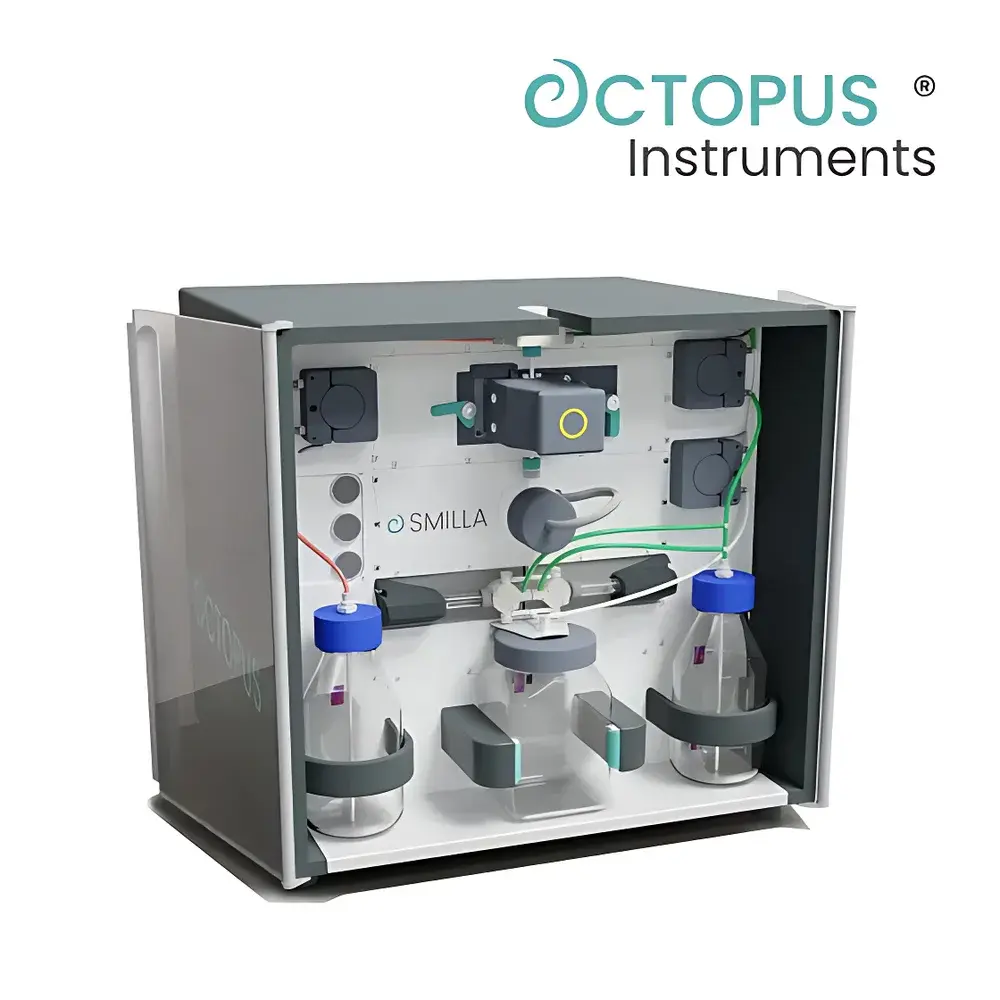
Octopus SAMILLA FIM FL High-Resolution Flow Imaging Microscopy System
| Brand | Octopus |
|---|---|
| Origin | Germany |
| Model | SAMILLA FIM FL |
| Camera Resolution | 12 MP |
| Magnification | 40× |
| Particle Detection Principle | Morphological Image Analysis |
| Measurable Size Range | 0.2 µm – 2 mm |
| Imaging Modes | Transmission Light (standard), Reflectance Light (optional) |
| Compliance | USP <788>, USP <789>, Ph. Eur. 2.9.19, ISO 21501-4, FDA 21 CFR Part 11 |
| Software | SMILLA View with Audit Trail, LIMS Integration, and GLP/GMP Workflow Support |
Overview
The Octopus SAMILLA FIM FL is a modular, high-resolution flow imaging microscopy (HiRes-FIM) system engineered for quantitative morphological analysis of suspended particles in liquid media. It operates on the principle of hydrodynamic focusing combined with real-time digital imaging—capturing high-fidelity, in-focus micrographs of individual particles as they pass through a precisely defined optical detection zone at controlled laminar flow. Unlike conventional light obscuration or static microscopy methods, HiRes-FIM delivers both dimensional metrics (length, width, aspect ratio, circularity, convexity) and visual confirmation of particle identity—including biological morphology (e.g., algal cell structure, diatom frustules, cyanobacterial filaments) and non-biological contaminants (e.g., fibers, silicone droplets, polymer fragments). Designed for regulatory-grade applications in environmental monitoring, pharmaceutical water quality control, and aquatic ecology research, the SAMILLA FIM FL meets stringent requirements for traceability, reproducibility, and data integrity under GLP and GMP frameworks.
Key Features
- Modular architecture supporting four interchangeable functional units: HiRes-FIM Imaging Module, Light Obscuration (LO) Module, Precision Pump Module (MACRO/MICRO1/MICRO2/HYBRID configurations), and Auto-Clean Module—all auto-recognized via embedded RFID tags upon insertion.
- 12-megapixel monochrome or color CMOS camera with ultra-short exposure time (<1 µs), eliminating motion blur and enabling reliable imaging of particles down to 0.2 µm in diameter under ISO 21501-4-compliant conditions.
- Motorized, software-controlled zoom optics with rapid-switching magnification modules (including 40× objective), coupled with autofocus algorithms that eliminate reliance on calibration beads.
- Dual illumination capability: standard brightfield transmission mode for transparent or pigmented planktonic organisms; optional reflectance mode for optically dense or highly scattering particles.
- Integrated fluidic intelligence: pressure sensors, flow meters, and temperature monitors ensure consistent hydraulic performance across varying sample viscosities and particulate loadings.
Sample Compatibility & Compliance
The SAMILLA FIM FL accepts aqueous samples ranging from raw surface water and wastewater effluents to purified pharmaceutical waters (PW, WFI) and algal culture suspensions. Its wide dynamic size range—from submicron colloids (200 nm) to macroplankton (>2 mm)—supports standardized enumeration of phytoplankton, zooplankton, and detrital aggregates per EN 16695, ISO 13395, and EPA Method 180.1. The system fully complies with pharmacopeial standards for subvisible particle characterization, including USP , USP , and Ph. Eur. 2.9.19. All analytical workflows support 21 CFR Part 11–compliant electronic records, featuring role-based user access, full audit trail logging, electronic signatures, and immutable data archiving—validated for use in regulated QC/QA laboratories subject to FDA, EMA, or PMDA inspection.
Software & Data Management
SMILLA View is a purpose-built, Windows-native application delivering synchronized control of all hardware modules and comprehensive image-based analytics. It provides pixel-accurate particle segmentation using adaptive thresholding and machine learning–enhanced classification models trained on reference morphological libraries (e.g., diatoms, dinoflagellates, green algae). Each detected object is assigned a unique ID linked to its raw image, centroid coordinates, geometric descriptors, and temporal acquisition stamp. Advanced filtering enables multi-parameter queries (e.g., “all elongated particles >15 µm with chlorophyll autofluorescence signature”), while batch processing supports statistical reporting aligned with ISO 5725 precision criteria. Data export formats include CSV, PDF, and XML; native integration with LIMS via ASTM E1384-compliant interfaces ensures seamless transfer into enterprise laboratory informatics ecosystems. Customizable report templates meet internal SOPs and external submission requirements (e.g., regulatory filings, inter-laboratory proficiency testing).
Applications
- Environmental water quality assessment: taxonomic identification and abundance quantification of phytoplankton and zooplankton communities in lakes, rivers, reservoirs, and coastal zones.
- Drinking water and wastewater treatment optimization: real-time monitoring of floc morphology, biofilm detachment events, and filter breakthrough indicators.
- Pharmaceutical manufacturing: routine subvisible particle testing of process water, final drug product solutions, and container-closure integrity verification.
- Aquaculture and algal biotechnology: viability assessment, growth stage classification, and contamination screening in photobioreactor cultures.
- Microplastic research: morphometric profiling of synthetic fibers, fragments, and beads—including shape-based differentiation from natural organic particulates.
FAQ
What particle size range can the SAMILLA FIM FL resolve with morphological fidelity?
It reliably images and classifies particles from 0.2 µm to 2 mm, with optimal morphological detail achieved between 1 µm and 500 µm using the 40× HiRes-FIM module.
Does the system require daily recalibration or focus-bead alignment?
No—autofocus is performed continuously during acquisition using contrast-based algorithms; no physical focus beads or manual intervention is needed.
Can SMILLA View distinguish live from dead algal cells?
Yes, when paired with optional fluorescence excitation modules (e.g., 488 nm LED), it supports chlorophyll-a autofluorescence intensity mapping and spectral ratio analysis for physiological status inference.
Is method validation support available for regulatory submissions?
Octopus provides IQ/OQ/PQ documentation packages, validation protocols aligned with ICH Q5A(R2) and USP , and on-site qualification assistance by certified field application scientists.
How does the Auto-Clean Module prevent carryover between high-concentration samples?
It executes a three-stage protocol: post-run flush with deionized water, reverse-flow purge to dislodge adherent particles, and air-drying of the flow cell—fully automated and logged within the audit trail.
Related Products