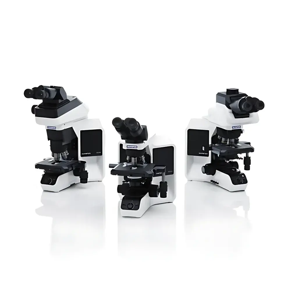
Olympus BX53(LED)/BX43/BX46 Clinical-Grade Upright Microscope
| Brand | Olympus |
|---|---|
| Origin | Japan |
| Manufacturer Type | Original Equipment Manufacturer (OEM) |
| Origin Category | Imported Instrument |
| Model Series | BX53(LED), BX43, BX46 |
| Instrument Type | Upright Microscope |
| Illumination Source | High-CRI White LED |
| Design Focus | Ergonomic Clinical Workflow Optimization |
Overview
The Olympus BX53(LED), BX43, and BX46 represent a family of clinical-grade upright microscopes engineered for routine histopathology, cytology, hematology, and clinical laboratory diagnostics. These instruments operate on conventional brightfield, phase contrast, and optional polarized light modalities—leveraging Olympus’ proprietary UIS2 optical system to deliver high numerical aperture (NA), chromatic aberration-corrected imaging across the visible spectrum (400–700 nm). Designed specifically for high-throughput clinical environments, the BX series integrates mechanical stability, thermal management for long-duration LED illumination, and modular optical pathways compatible with digital camera adapters, fluorescence filter cubes (when equipped), and motorized stage options. Unlike research-oriented inverted platforms, the upright configuration enables rapid slide handling, direct immersion oil application, and seamless integration into existing clinical workflow benches without requiring reconfiguration of specimen orientation or operator posture.
Key Features
- Ergonomic optical train with adjustable interpupillary distance (55–75 mm), inclined binocular tube (0–30° tilt), and low-positioned focus knobs—reducing cervical strain during extended diagnostic sessions.
- Long-life, flicker-free white LED illumination (6,000 K CCT, >90 CRI) providing stable luminance (>1,800 cd/m² at field stop) with no warm-up time, minimal heat emission (<1.2 W thermal load at objective collar), and compliance with IEC 62471 photobiological safety standards.
- Modular UIS2 infinity-corrected optics supporting 4× to 100× objective magnifications, including semi-apochromatic and apochromatic objectives optimized for consistent color fidelity and edge-to-edge flatness (≤±0.02% field curvature).
- Robust mechanical stage with dual-scale vernier readout (0.1 mm resolution), spring-loaded specimen clips, and optional motorized XY control (BX53 only) compliant with RS-232 and USB 2.0 host interfaces.
- Interchangeable observation tubes (trinocular, ergonomic binocular, high-eyepoint wide-field) and standardized dovetail-mounted condenser systems (Abbe, achromatic-aplanatic, or phase slider-compatible) enabling rapid adaptation to ISO 10993-5 cytotoxicity slide screening or CLIA-certified Pap smear analysis protocols.
Sample Compatibility & Compliance
The BX series accommodates standard 1″ × 3″ glass microscope slides (thickness: 0.9–1.2 mm) and 22 × 22 mm coverslips (0.13–0.17 mm), with objective working distances calibrated per ISO 8578 for dry (≥0.55 mm WD at 40×) and oil-immersion (≥0.13 mm WD at 100×) configurations. All models meet CE marking requirements under Directive 2014/30/EU (EMC) and 2014/35/EU (LVD), and are classified as Class I medical devices per MDR (EU) 2017/745 when deployed in diagnostic pathology workflows. The BX53(LED) variant supports optional FDA 21 CFR Part 11–compliant digital acquisition modules (e.g., DP80 camera + cellSens software) with audit trail logging, electronic signature enforcement, and role-based user access controls aligned with CAP and COLA laboratory accreditation criteria.
Software & Data Management
When paired with Olympus’ cellSens Dimension software (v3.1+), the BX platform enables DICOM-compliant image acquisition, multi-layer Z-stack reconstruction, and quantitative morphometric analysis (nuclear area, cytoplasmic ratio, mitotic index scoring) validated per CAP Q-Probes benchmarks. Raw TIFF and JPEG2000 export formats preserve bit-depth fidelity (12-bit dynamic range), while metadata embedding includes objective ID, magnification, exposure time, LED intensity setting, and operator-assigned case ID—all traceable via embedded EXIF tags. For LIS/HIS integration, HL7 v2.5.1 message routing is supported through optional middleware gateways, ensuring bidirectional synchronization of accession numbers, patient demographics, and annotation timestamps without manual re-entry.
Applications
- Routine histopathological evaluation of H&E-, IHC-, and special-stain–processed tissue sections in hospital-based surgical pathology labs.
- Cytological screening of liquid-based Papanicolaou (Pap) smears and fine-needle aspiration (FNA) specimens under standardized Bethesda System criteria.
- Hematology differential counts using Wright-Giemsa–stained peripheral blood films, with objective-specific calibration for erythrocyte size distribution and platelet morphology assessment.
- Microbiological identification of Gram-stained bacterial isolates and acid-fast bacilli (AFB) using high-NA oil objectives and Köhler-optimized condenser alignment.
- Quality assurance verification of immunohistochemistry staining uniformity and antigen retrieval efficacy in CAP-accredited reference laboratories.
FAQ
Is the BX53(LED) suitable for GLP-compliant histology documentation?
Yes—the BX53(LED) with DP80 camera and cellSens software supports full audit trail generation, electronic signatures, and secure user authentication required under OECD GLP Principles and FDA Guidance for Industry (2022).
Can BX43 and BX46 be upgraded to support fluorescence imaging?
No—only the BX53 platform offers integrated fluorescence capability via its universal filter turret and optional mercury or LED excitation sources; BX43/BX46 lack the requisite optical path symmetry and dichroic mirror mounts.
What maintenance intervals are recommended for clinical deployment?
Olympus recommends quarterly optical alignment verification (per ISO 10993-10), annual LED output calibration (using NIST-traceable photometer), and biannual cleaning of condenser lens elements using ISO 10110–7–compliant lint-free wipes and anhydrous ethanol.


