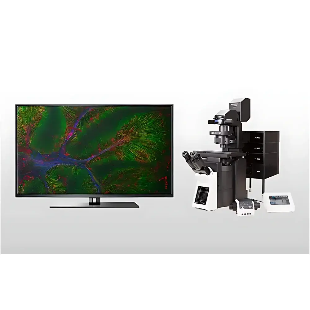
Olympus/EVIDENT FV4000 Laser Scanning Confocal Microscope
| Brand | Olympus/EVIDENT |
|---|---|
| Origin | Japan |
| Instrument Type | Point-Scanning Confocal Microscope |
| Laser Options | Multiple NIR Wavelengths (405–785 nm, up to 10 lines) |
| Detectors | Next-Generation Silicon Photomultiplier (SiPM)-Based SilVIR Detectors |
| Detection Channels | Up to 6 Simultaneous Fluorescence Channels |
| Spectral Imaging | TruSpectral Full-Spectrum Detection |
| Scan Unit | Hybrid Scanning Module (Galvo + Resonant Scanner) |
| Microscope Frame | Research-Grade Motorized Stand (Inverted, Upright, Electrophysiology, or Gantry Configurations) |
| Illumination Sources | LED-Based Fluorescence, Brightfield LED, or Halogen Lamp Options |
| Software Platform | Multi-Module Intelligent Control Suite with Experimental Workflow Design, Quantitative Image Management, and Audit-Trail-Capable Data Archiving |
Overview
The Olympus/EVIDENT FV4000 Laser Scanning Confocal Microscope is a high-performance point-scanning confocal system engineered for quantitative, multi-scale biological imaging. It operates on the fundamental principle of optical sectioning via focused laser excitation and pinhole-confined detection—enabling precise rejection of out-of-focus light and generation of high-contrast, z-resolved volumetric datasets. Designed for rigorous life science research—including live-cell dynamics, subcellular organelle tracking, tissue-level morphology, and multimodal correlative workflows—the FV4000 integrates advanced photodetection, spectral flexibility, and motorized precision into a modular platform compliant with GLP/GMP-aligned laboratory practices.
Key Features
- SilVIR Silicon Photomultiplier (SiPM) Detectors: Patented detector architecture combining high-gain SiPM sensor elements with ultra-low-noise, high-bandwidth signal processing—delivering >90% quantum efficiency in the visible-to-NIR range (405–785 nm), minimal sensitivity drift over time, and linear photon-counting response across six independent detection channels.
- TruSpectral Full-Spectrum Detection: Enables simultaneous acquisition of spectral fluorescence data without filter wheel switching—supporting unmixing of spectrally overlapping fluorophores and quantitative spectral fingerprinting with sub-nanometer resolution.
- Hybrid Scanning Architecture: Combines galvanometric mirrors for high-fidelity, low-distortion imaging with resonant scanners for high-speed frame rates (up to 30 fps at 512 × 512), facilitating time-lapse studies of dynamic cellular processes under physiological conditions.
- Multi-Configuration Motorized Stand: Supports inverted, upright, electrophysiology-integrated, and gantry-style configurations—all fully automated for objective turret, filter cube, stage, focus, and Z-drift compensation (TruFocus real-time feedback loop).
- Expanded Laser Excitation Range: Up to ten discrete laser lines spanning 405 nm to 785 nm—including multiple NIR options—optimized for deep-tissue penetration, reduced phototoxicity, and compatibility with far-red and NIR-II probes.
- Laser Power Monitoring (LPM): Integrated real-time power calibration ensures consistent photon flux across sessions, operators, and instruments—critical for longitudinal quantification and inter-laboratory reproducibility.
Sample Compatibility & Compliance
The FV4000 accommodates diverse specimen formats—from adherent monolayers and 3D organoids to thick tissue sections (up to 500 µm with NIR optimization) and whole-mount preparations. Its optical design complies with ISO 10993-5 (biocompatibility of optical components in contact with biological samples) and supports FDA 21 CFR Part 11–compliant software operation when configured with audit-trail-enabled user authentication, electronic signatures, and immutable metadata logging. All hardware modules meet IEC 61000-6-3 EMC emission standards and IEC 61000-6-2 immunity requirements for laboratory environments.
Software & Data Management
FV4000 is controlled by Olympus’ multi-module intelligent software suite—designed for complex experimental orchestration, including multi-position time-lapse, multi-channel spectral acquisition, and automated Z-stack tiling. The platform supports DICOM-SR export for clinical correlation, OME-TIFF standardization for FAIR data principles, and hierarchical project-based archiving with embedded acquisition metadata (laser power, dwell time, pinhole size, gain, offset, objective ID). Raw photon-counting data is preserved in HDF5 format, enabling post-acquisition spectral unmixing, deconvolution, and machine learning–ready dataset curation. Software validation documentation is available per IQ/OQ protocols for regulated environments.
Applications
- Quantitative co-localization analysis of protein complexes using spectral unmixing and Pearson’s correlation coefficient computation.
- Long-term live-cell imaging of calcium dynamics, mitochondrial membrane potential, and vesicular trafficking with minimized photobleaching.
- 3D reconstruction and morphometric analysis of neuronal dendritic spines, glomerular tufts, or tumor vasculature.
- Correlative light-electron microscopy (CLEM) workflows leveraging precise coordinate mapping and fiducial marker registration.
- Preclinical imaging of NIR-labeled antibodies, nanoparticles, or reporter constructs in ex vivo tissue slices and cleared organs.
- Standardized assay development for high-content screening (HCS) where inter-instrument comparability of intensity values (photons/pixel) is required.
FAQ
What laser wavelengths are supported on the FV4000?
The system offers up to ten factory-installed laser lines ranging from 405 nm to 785 nm, including optional NIR lasers at 730 nm and 785 nm for deep-tissue applications.
Can the FV4000 be upgraded to multiphoton capability?
Yes—the modular architecture supports field-installable upgrade paths to integrated multiphoton excitation via external femtosecond laser coupling and non-descanned detection pathways.
Is the software compliant with 21 CFR Part 11 requirements?
When deployed with validated configuration, electronic signature modules, and audit-trail logging enabled, the software meets core technical controls outlined in FDA 21 CFR Part 11 for electronic records and signatures.
How does SilVIR improve quantitative reproducibility compared to PMT-based systems?
SilVIR detectors exhibit <0.5% sensitivity drift over 12 months and linear response across 6 decades of photon flux—eliminating gain recalibration needs and enabling cross-platform intensity normalization using absolute photon counts.
Does the FV4000 support automated Z-drift correction during long acquisitions?
Yes—TruFocus uses real-time infrared reflection feedback from the coverslip interface to dynamically adjust objective position with sub-10 nm stability over multi-hour time-lapse sessions.


