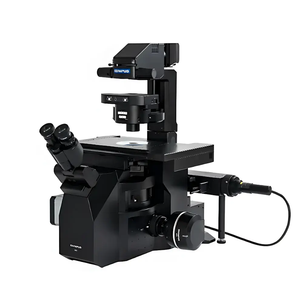
Olympus IXplore IX85 Fully Motorized Inverted Microscope
| Brand | Olympus |
|---|---|
| Origin | Japan |
| Manufacturer Type | Original Equipment Manufacturer (OEM) |
| Origin Category | Imported |
| Model | IXplore IX85 |
| Instrument Type | Inverted Microscope |
| Field Number (FN) | 26.5 mm |
| Optical Design | Widefield, Plan Apochromat-Corrected Optics with Distortion-Free Imaging |
| Immersion Medium Support | Silicone Oil Immersion (Auto-Tracking Silicon Gel), Water, Glycerol, Oil |
| Correction Collar | Motorized, Auto-Adjusting for Coverslip Thickness (0.13–0.22 mm) |
| Dual Port Architecture | Integrated Dual Trinocular Ports (Simultaneous Widefield + Confocal or TIRF Pathways) |
| Modular Expansion | Compatible with Spinning Disk Confocal, TIRF, Super-Resolution (e.g., SIM), and Optogenetic Manipulation Modules |
| Software Platform | cellSens Dimension v3.x with Real-Time Deconvolution, Stitching, and GLP-Compliant Audit Trail (21 CFR Part 11 Ready) |
Overview
The Olympus IXplore IX85 is a fully motorized inverted microscope platform engineered for high-content, multi-modal live-cell and tissue imaging in demanding life science laboratories. Built upon a rigid, thermally stable optical chassis, the system implements advanced Köhler illumination with uniform intensity distribution across its industry-leading 26.5 mm field number — enabling acquisition of large-area, distortion-free images without peripheral falloff or geometric aberration. Its optical path integrates Olympus’ proprietary Plan Apochromat objectives with extended flat-field correction and built-in distortion compensation algorithms, ensuring sub-micron spatial fidelity from center to edge. The platform operates on a dual-port architecture with independent, synchronized optical pathways — supporting simultaneous widefield fluorescence, transmitted light, and secondary modalities such as TIRF or spinning-disk confocal without mechanical realignment. Designed for longitudinal studies under physiological conditions, the IX85 accommodates environmental chambers (temperature, CO₂, humidity control) and integrates seamlessly with motorized stage systems for automated time-lapse and multi-position experiments.
Key Features
- Fully motorized operation: All critical components — focus, filter turrets, condenser, objective nosepiece, and XY stage — are precision-stepped and software-synchronized for reproducible, hands-free workflows.
- Silicone oil immersion optics: Proprietary silicone gel medium maintains refractive index stability (n = 1.40) across temperature fluctuations; auto-tracking gel delivery eliminates manual reapplication and minimizes spherical aberration at depth.
- Motorized correction collar: Dynamically adjusts objective correction ring position based on coverslip thickness (0.13–0.22 mm), compensating for spherical aberration in real time during Z-stack acquisition.
- Widefield imaging optimization: 26.5 mm field number increases single-frame coverage by ~44% compared to conventional 22 mm FN systems, reducing required tile count by up to 50% for large-area mosaics.
- Smart shading correction: Pixel-level illumination normalization applied in real time, enabling seamless stitching of >100-tile mosaics with <0.5% intensity variance across stitched boundaries.
- Open modular architecture: Standardized rail interface and power/data bus support plug-and-play integration of third-party or Olympus-developed modules including TIRF illuminators, spinning-disk confocal units, and optogenetic stimulation devices.
Sample Compatibility & Compliance
The IXplore IX85 supports diverse biological specimens — from adherent mammalian cells cultured on glass-bottom dishes to thick 3D organoids, zebrafish embryos, and ex vivo tissue slices up to 500 µm in thickness. Its long-working-distance objectives (e.g., UPLSAPO 20×/0.75 WD 3.1 mm) and adjustable condenser height accommodate variable sample heights and chamber geometries. The system complies with ISO 9001 manufacturing standards and meets CE marking requirements for laboratory instrumentation. When configured with cellSens Dimension v3.x and enabled audit trail logging, it satisfies documentation requirements for GLP and GMP environments per FDA 21 CFR Part 11 (electronic records and signatures). All optical components adhere to JIS B 7131 and ISO 10934-1 specifications for microscope performance verification.
Software & Data Management
Olympus cellSens Dimension v3.x serves as the unified control and analysis environment, offering integrated hardware orchestration, real-time image processing, and structured data export. Key capabilities include: multi-channel time-lapse scheduling with drift correction; GPU-accelerated deconvolution (Wiener and constrained iterative algorithms); AI-assisted segmentation using pre-trained models for nuclei, membranes, and organelles; and batch processing pipelines compliant with FAIR data principles. All user actions — parameter changes, acquisition triggers, and analysis steps — are logged with timestamps, operator ID, and system state metadata. Export formats include OME-TIFF (with full metadata embedding), HDF5, and standardized JSON-LD for LIMS integration. Remote monitoring and collaborative review are supported via secure HTTPS-based cellSens Web Viewer.
Applications
- High-throughput phenotypic screening in drug discovery, leveraging automated multi-well plate imaging and quantitative morphometric analysis.
- Long-term live-cell dynamics: mitosis tracking, intracellular trafficking, calcium signaling, and mitochondrial fission/fusion events under controlled environmental conditions.
- Multi-modal correlative imaging: sequential or simultaneous acquisition across widefield, TIRF, and spinning-disk confocal channels to resolve structural hierarchy from membrane nanodomains to whole-cell organization.
- 3D tissue imaging: optical sectioning of spheroids and organoids with motorized Z-control and adaptive focus stabilization (ZDC).
- Optogenetics-integrated experiments: precise spatiotemporal light delivery combined with real-time fluorescence readout using synchronized LED/laser excitation modules.
- Preclinical imaging validation: alignment with histopathology workflows through standardized coordinate mapping and digital slide export.
FAQ
What immersion media are supported by the IXplore IX85 objectives?
The system natively supports silicone oil (n = 1.40), water (n = 1.33), glycerol (n = 1.47), and standard immersion oil (n = 1.51), with dedicated objective series optimized for each medium.
Can the IX85 be upgraded to super-resolution capability post-purchase?
Yes — the open optical architecture and standardized mounting interfaces allow field-installation of structured illumination microscopy (SIM) modules, provided the base system includes compatible laser combiner and EMCCD/sCMOS detection paths.
Is the motorized correction collar compatible with all IX85 objectives?
Motorized correction is available on all Plan Apochromat objectives with correction collar designation (e.g., UPLSAPO 40×/0.95, UPLSAPO 60×/1.35), but not on fixed-collimation objectives such as UPlanSApo 100×/1.40 Oil.
Does cellSens Dimension support automated image analysis for publication-grade figures?
Yes — the software includes batch measurement tools compliant with MIAME and MIAPE reporting guidelines, with export options for TIFF (8/16-bit), SVG vector overlays, and annotated PDFs suitable for journal submission.
How does the IX85 handle thermal drift during extended time-lapse experiments?
The system incorporates a passive thermal stabilization chassis and optional active Z-drift compensation (ZDC) module that monitors focus position via infrared reflection and adjusts objective position at 10 Hz resolution to maintain sub-100 nm axial stability over 24+ hours.


