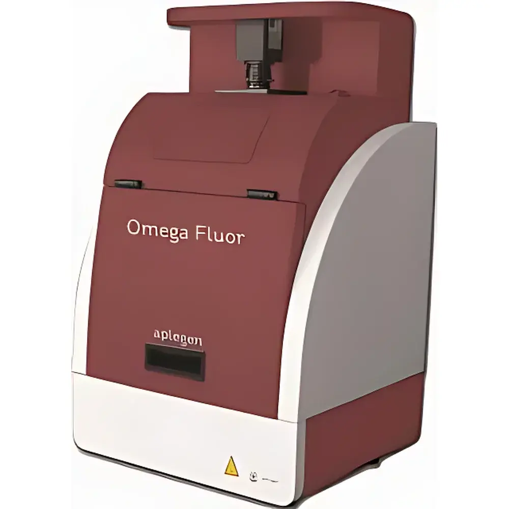
Omega Fluor Gel Imaging System
| Brand | Aplegen |
|---|---|
| Origin | USA |
| Manufacturer Type | Authorized Distributor |
| Instrument Category | Imported |
| Model | Omega Fluor |
| Instrument Type | Standard Gel Imaging System |
| CCD Resolution | 5.0 Megapixel (Physical Pixels) |
| Bit Depth | 12-bit |
| Lens | Fixed F1.4 Lens |
| Dimensions (W×D×H) | 36 × 32 × 64 cm |
| Safety Feature | UV Interlock — Automatic Power Cutoff Upon Chamber Door Opening |
| Compatible Stains | Ethidium Bromide (EB), Silver Stain, Coomassie Blue |
Overview
The Omega Fluor Gel Imaging System is a compact, high-performance benchtop instrument engineered for reliable, reproducible documentation of nucleic acid and protein gels under ultraviolet (UV) and visible light excitation. Designed around a scientific-grade 5.0-megapixel monochrome CCD sensor with true physical pixel architecture and 12-bit analog-to-digital conversion, the system delivers exceptional dynamic range and low-noise image acquisition—critical for quantitative band intensity analysis in molecular biology workflows. Its optical path integrates a fixed F1.4 wide-aperture lens optimized for maximum photon capture across UV (254 nm and 302 nm) and white-light illumination modes, eliminating focus drift and mechanical variability associated with motorized or manual focusing mechanisms. The system operates on the principle of fluorescence imaging: UV radiation excites intercalating dyes (e.g., ethidium bromide) bound to DNA, while visible-light transillumination or reflected illumination enables visualization of Coomassie- or silver-stained protein gels. All illumination sources are housed within a fully shielded, interlocked dark chamber compliant with IEC 61010-1 safety standards.
Key Features
- True 5.0-megapixel CCD sensor with 12-bit digitization ensures high-fidelity capture of fine electrophoretic band detail and accurate grayscale linearity across four orders of magnitude.
- F1.4 fixed-focus lens provides consistent optical performance without calibration drift—ideal for routine lab use where speed and repeatability supersede variable magnification needs.
- Integrated UV safety interlock: Chamber door sensors immediately terminate UV lamp power upon opening, meeting OSHA and ISO 15223-1 labeling requirements for Class 3R UV devices.
- Benchtop footprint (36 × 32 × 64 cm) minimizes spatial demand while accommodating standard mini- and midi-gel formats (up to 15 × 15 cm).
- Dual-mode illumination: Independent 302 nm UV transilluminator for EB-stained DNA gels; adjustable white LED array for Coomassie- and silver-stained SDS-PAGE gels.
- No software dongles or hardware keys required—system firmware and acquisition software are pre-installed and licensed per unit.
Sample Compatibility & Compliance
The Omega Fluor supports standard slab gel formats used in agarose and polyacrylamide electrophoresis, including mini-gels (8 × 10 cm), midi-gels (10 × 12 cm), and large-format gels up to 15 × 15 cm. It accommodates both horizontal (agarose) and vertical (SDS-PAGE) configurations without accessory reconfiguration. Stain compatibility includes ethidium bromide (EB), SYBR® Safe, GelRed®, silver nitrate-based protocols (e.g., Blum method), and Coomassie Brilliant Blue R-250/G-250. The system conforms to key regulatory expectations for documentation integrity: image metadata (exposure time, lens aperture, illumination mode, timestamp) is embedded in TIFF headers; raw acquisition files are stored unaltered, supporting audit trails required under GLP and FDA 21 CFR Part 11–aligned laboratory practices when paired with validated LIMS integration.
Software & Data Management
Acquisition and basic analysis are performed via Aplegen’s proprietary OmegaCapture software (v4.x), compatible with Windows 10/11 (64-bit). The interface enables one-click capture, real-time histogram preview, auto-exposure optimization, and batch processing of multi-gel series. Export options include lossless TIFF (12-bit), PNG, and PDF with embedded scale bars and annotation layers. Quantitative tools include lane profiling, molecular weight estimation using reference ladders, and relative band intensity normalization against internal controls. Raw image files retain full bit-depth fidelity and are structured in a hierarchical folder system compliant with FAIR data principles (Findable, Accessible, Interoperable, Reusable). No cloud dependency—data remains on local storage or network-attached drives per institutional IT policy.
Applications
- Documentation and semi-quantitative analysis of PCR products, restriction digests, and DNA laddering in agarose gels.
- Post-electrophoretic validation of CRISPR editing efficiency via T7E1 or Surveyor assays.
- Quality control of purified plasmid preps and genomic DNA extractions.
- Protein expression profiling across cell lysates, purification fractions, or time-course experiments using Coomassie- or silver-stained SDS-PAGE.
- Teaching laboratory use: intuitive interface and rapid workflow reduce training overhead for undergraduate and graduate students.
FAQ
Is the Omega Fluor compatible with SYBR® Gold or other next-generation nucleic acid stains?
Yes—the 302 nm UV transilluminator provides optimal excitation for SYBR® Gold, SYBR® Green I, and GelGreen®. Emission filters are not required for standard documentation but may be added as optional accessories for enhanced signal-to-noise in low-abundance samples.
Can I perform densitometry and publish normalized band intensities from OmegaCapture output?
Yes—OmegaCapture computes integrated density values (IDV) per band using background-subtracted pixel summation. When calibrated against known standards and acquired under consistent exposure conditions, results meet the technical rigor expected for publication in journals requiring adherence to MIQE or MIAPE guidelines.
Does the system support time-lapse imaging for in-gel enzymatic assays?
No—the Omega Fluor is designed for endpoint imaging only. It does not offer programmable multi-frame acquisition or temperature-controlled gel stages required for kinetic assays.
What maintenance is required for long-term optical stability?
Annual verification of UV lamp output intensity (using NIST-traceable radiometer) and lens cleanliness is recommended. No alignment or recalibration procedures are necessary due to the fixed-optics architecture.


