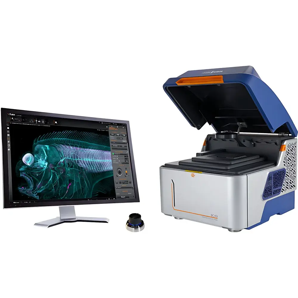
Oxford Instruments ANDOR BC43 Benchtop Confocal Microscope
| Brand | Oxford Instruments |
|---|---|
| Origin | United Kingdom |
| Model | BC43 |
| Imaging Modes | Confocal, Widefield, Brightfield & Differential Phase Contrast (DPC) Transmission |
| Illumination Technology | Patented Borealis™ Uniform Illumination |
| Z-Stack Speed | ≥10× faster than point-scanning confocal systems |
| Software Platform | ANDOR Fusion (GPU-accelerated deconvolution, multi-position montage, hardware-synchronized 3D stitching) |
| Compliance | Designed for GLP/GMP-aligned workflows |
| Physical Design | Compact benchtop architecture with integrated vibration damping and optical sealing — no darkroom or optical table required |
Overview
The Oxford Instruments ANDOR BC43 is a high-performance benchtop confocal microscope engineered for quantitative, multi-modal life science imaging. Unlike traditional point-scanning confocal systems that rely on sequential laser spot rasterization — a process inherently limited by photobleaching, acquisition speed, and signal-to-noise ratio — the BC43 employs a spinning-disk-based parallel confocal architecture combined with ANDOR’s proprietary Borealis™ uniform illumination technology. This design enables simultaneous excitation and detection across thousands of spatial points, delivering true optical sectioning at speeds exceeding those of conventional confocal platforms by over an order of magnitude. The system supports three complementary imaging modalities: confocal fluorescence for high-contrast, background-free 3D reconstruction; widefield fluorescence with GPU-accelerated deconvolution for rapid, high-sensitivity acquisition in thin specimens; and dual-mode transmission imaging (brightfield and Differential Phase Contrast, DPC) for label-free structural visualization. Its compact, vibration-isolated benchtop form factor — featuring an optically sealed enclosure and ergonomic joystick-controlled stage — eliminates dependency on dedicated optical tables or darkrooms while maintaining sub-micron axial resolution and exceptional photostability across extended time-lapse experiments.
Key Features
- Parallel confocal acquisition engine delivering ≥10× faster volumetric imaging versus point-scanning systems, without compromising lateral or axial resolution
- Borealis™ illumination technology ensuring >98% intensity uniformity across FOV — critical for quantitative intensity measurements and seamless tile-based montage stitching
- Integrated multi-modal platform supporting confocal, widefield, brightfield, and DPC transmission imaging within a single instrument
- Ergonomic joystick interface with 2× overview objective for rapid sample navigation and region-of-interest selection
- Benchtop mechanical architecture with passive vibration damping and hermetic optical sealing — operational stability verified under standard laboratory ambient conditions
- Fusion software suite with hardware-synchronized multi-position acquisition, real-time GPU-accelerated deconvolution, and automated 3D montage stitching
Sample Compatibility & Compliance
The BC43 accommodates diverse biological specimens ranging from subcellular structures (e.g., microtubule dynamics, synaptic vesicle trafficking) to intact cleared tissues and live model organisms up to 5 mm in thickness. Its long-working-distance objectives support imaging depths exceeding 300 µm in cleared zebrafish embryos and Drosophila larvae, while DPC mode enables high-contrast visualization of unstained, low-contrast samples such as early-stage organoids or translucent hydrogel-embedded constructs. The system complies with international standards relevant to regulated research environments: image metadata conforms to OME-TIFF specifications; raw data export supports Imaris IMS format for downstream analysis in ISO/IEC 17025- or GLP-compliant pipelines; and when integrated with validated LIMS and electronic lab notebook (ELN) systems, the BC43 supports audit-trail generation aligned with FDA 21 CFR Part 11 requirements.
Software & Data Management
ANDOR Fusion serves as the unified control and analysis interface for the BC43. It provides intuitive workflow templates for multi-dimensional acquisitions — including time-lapse Z-stacks, multi-position montages, and multi-channel spectral unmixing — all configurable via drag-and-drop protocols. Real-time GPU-accelerated deconvolution (Richardson-Lucy algorithm) enhances widefield image resolution without post-processing latency. Hardware synchronization ensures pixel-perfect alignment between stage movement, focus drive, and camera exposure during large-area tiled acquisitions. All acquired datasets are stored in open-format OME-TIFF containers with embedded metadata (objective magnification, emission filter set, Z-step size, laser power), enabling reproducible reprocessing and FAIR (Findable, Accessible, Interoperable, Reusable) data management. Native Imaris IMS export facilitates advanced surface rendering, colocalization quantification, and 4D trajectory analysis — particularly valuable for developmental biology, neuroimaging, and tumor spheroid studies.
Applications
- Developmental Biology: High-speed, low-photoxicity imaging of live zebrafish and Drosophila embryos through gastrulation and organogenesis; multi-region montage acquisition across entire specimens with micron-level registration accuracy
- Cell Biology: Quantitative tracking of subcellular organelles over >24-hour periods; simultaneous visualization of nm-scale cytoskeletal elements and mm-scale tissue architecture in 3D culture models
- Tissue Imaging: Confocal-grade optical sectioning in chemically cleared mouse brain sections or human tumor biopsies; seamless stitching of >50-tile montages covering whole-organ cross-sections at cellular resolution
- Organoid & 3D Culture Research: Long-term viability-preserving imaging of intestinal, cerebral, or pancreatic organoids under physiological conditions; dynamic assessment of lumen formation, cell polarization, and barrier integrity
FAQ
Does the BC43 require a dedicated darkroom or optical table?
No. Its fully enclosed, vibration-damped benchtop chassis and internal optical sealing enable stable operation in standard laboratory environments.
Can the BC43 perform true optical sectioning without computational deconvolution?
Yes. Its spinning-disk confocal architecture provides inherent optical sectioning capability — deconvolution is optional and used only to enhance widefield data.
Is Imaris included with the BC43, or is it licensed separately?
Imaris is available as an optional add-on module; the base system includes full Fusion software with native IMS export compatibility.
What level of training is required to operate the BC43 independently?
Minimal. The joystick-driven interface and preconfigured acquisition workflows allow new users to acquire publication-quality images within one training session.
How does the BC43 handle phototoxicity during live-cell imaging?
By minimizing total illumination dose through parallel detection and ultrafast exposure control, the BC43 achieves sub-10 mW/mm² effective irradiance — well below thresholds known to induce measurable photodamage in mammalian cells.


