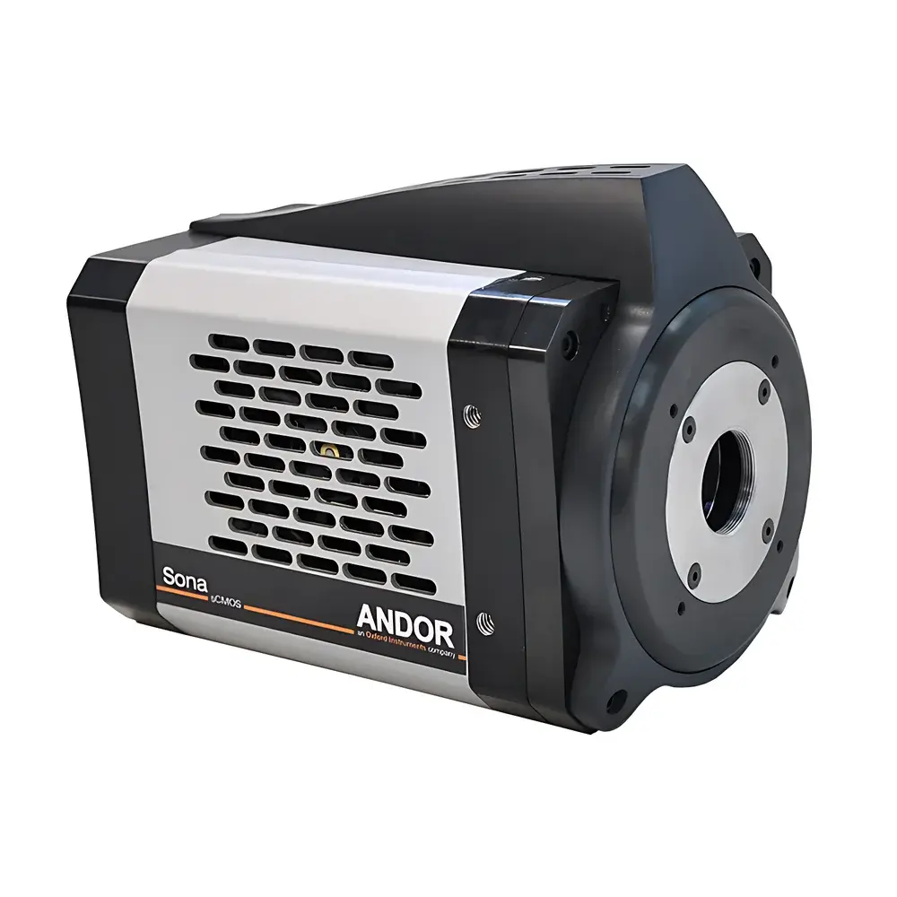
Oxford Instruments Andor Sona 4.2B-6 Back-Illuminated sCMOS Microscopy Camera Platform
| Brand | Oxford Instruments |
|---|---|
| Origin | United Kingdom |
| Model | Sona 4.2B-6 |
| Image Resolution | 2048 × 2048 |
| Pixel Size | 6.5 µm × 6.5 µm |
| Sensor Dimensions | 13.3 mm × 13.3 mm |
| Onboard Memory | 1 GB |
| Readout Speed | 310 MHz |
| Dynamic Range | 34,000:1 |
| Quantum Efficiency | 95% |
Overview
The Oxford Instruments Andor Sona 4.2B-6 is a vacuum-cooled, back-illuminated scientific CMOS (sCMOS) camera engineered for quantitative fluorescence microscopy in life science applications. It operates on the fundamental principle of photon-limited imaging—maximizing signal capture while minimizing read noise, dark current, and fixed-pattern artifacts. The camera employs a monolithic, back-thinned sCMOS sensor sealed within an ultra-high-vacuum chamber and actively cooled to –45 °C, enabling sub-electron read noise performance and thermal dark current suppression below 0.001 e–/pix/s. This architecture ensures high-fidelity acquisition across demanding modalities including confocal, TIRF, light-sheet, and super-resolution microscopy—where photon budget, temporal resolution, and spatial fidelity are simultaneously constrained.
Key Features
- Back-illuminated sCMOS sensor with 95% peak quantum efficiency (QE) at 560 nm, delivering industry-leading photon detection capability for low-light biological specimens.
- 2048 × 2048 pixel array with 6.5 µm pixel pitch—optimized for Nyquist sampling with high-numerical-aperture (NA ≥ 1.3) 40× and 60× objectives on standard microscope C-mount interfaces.
- Vacuum-sealed, thermoelectrically cooled architecture maintaining stable –45 °C sensor temperature without condensation or maintenance, ensuring long-term calibration stability and reproducibility.
- CoaXPress 2.0 interface supporting full-frame 16-bit image acquisition at up to 74 fps—scalable to >500 fps in user-defined ROI modes without compromising bit depth or linearity.
- Low-noise acquisition mode that reduces system read noise by >30% relative to standard operation—without increasing exposure time or sacrificing frame rate—enabling robust single-molecule tracking and fast Ca2+ dynamics imaging.
- 1 GB onboard frame buffer for sustained burst acquisition and real-time buffering during high-throughput time-lapse or multi-channel experiments.
Sample Compatibility & Compliance
The Sona 4.2B-6 is designed for integration into regulated and non-regulated research environments. Its hardware design and firmware support compliance with ISO/IEC 17025 traceability requirements for measurement uncertainty reporting. While not a medical device, its performance characteristics align with best practices outlined in ASTM E2717 (Standard Practice for Evaluating Performance of Digital Imaging Systems in Microscopy) and ISO 10934-1 (Optics and optical instruments — Vocabulary for microscopy). For GLP/GMP-aligned labs, the camera’s deterministic timing, hardware-triggered acquisition, and metadata-rich TIFF output facilitate audit-ready documentation. Optional Andor SDK integration supports FDA 21 CFR Part 11-compliant software environments when deployed with validated third-party acquisition platforms.
Software & Data Management
The Sona 4.2B-6 is fully supported by Andor’s Fusion software suite—a modular, Python- and MATLAB-compatible platform offering real-time visualization, multi-dimensional acquisition scripting (Z-stacks, time-series, spectral unmixing), and automated calibration workflows. All images include embedded EXIF-style metadata: exposure time, gain, sensor temperature, timestamp (with microsecond precision), and hardware configuration state. Raw data is saved in lossless 16-bit TIFF format with optional HDF5 export for scalable analysis in tools such as ImageJ/Fiji, Python (scikit-image, napari), or MATLAB. Fusion also provides built-in flat-field correction, pixel defect mapping, and non-uniformity compensation—calibrated per unit during factory certification and field-upgradable via secure firmware updates.
Applications
- Live-cell fluorescence imaging requiring high spatiotemporal resolution under minimal illumination (e.g., GFP-tagged cytoskeletal dynamics, mitochondrial fission/fusion).
- Single-molecule localization microscopy (SMLM), including dSTORM and PALM, where high QE and low noise directly improve localization precision and labeling density.
- High-speed calcium imaging in neuronal cultures or brain slices using GCaMP variants—leveraging 74 fps full-frame throughput to resolve sub-100 ms transients.
- Multi-color, multi-plane acquisitions in spinning-disk confocal systems where inter-channel crosstalk and frame synchronization must be tightly controlled.
- Quantitative co-localization analysis and FRET efficiency mapping—enabled by linear response over 4.5 decades and <0.1% pixel-to-pixel gain variation.
FAQ
What cooling method does the Sona 4.2B-6 use, and how is thermal stability maintained?
The camera utilizes a two-stage thermoelectric cooler (TEC) integrated into a hermetically sealed, ultra-high-vacuum chamber. This eliminates condensation risk and ensures sensor temperature remains stable within ±0.1 °C over multi-hour acquisitions.
Is the Sona 4.2B-6 compatible with third-party microscope control software?
Yes—it supports standard communication protocols including GenICam v3.1, DCAM, and Andor’s open-source SDK (C++, Python, .NET), enabling seamless integration with Micro-Manager, MetaMorph, and custom LabVIEW or MATLAB applications.
How does the low-noise mode affect dynamic range and linearity?
Low-noise mode maintains full 16-bit linearity and preserves the specified 34,000:1 dynamic range by optimizing correlated double sampling (CDS) timing and analog gain staging—not by binning or clipping.
Can the camera be used in environments requiring electromagnetic compatibility (EMC) certification?
The Sona 4.2B-6 complies with EN 61326-1:2013 for laboratory equipment and meets radiated emission limits per CISPR 11 Group 1, Class B, making it suitable for shared core facility spaces adjacent to sensitive electrophysiology or mass spectrometry instrumentation.


