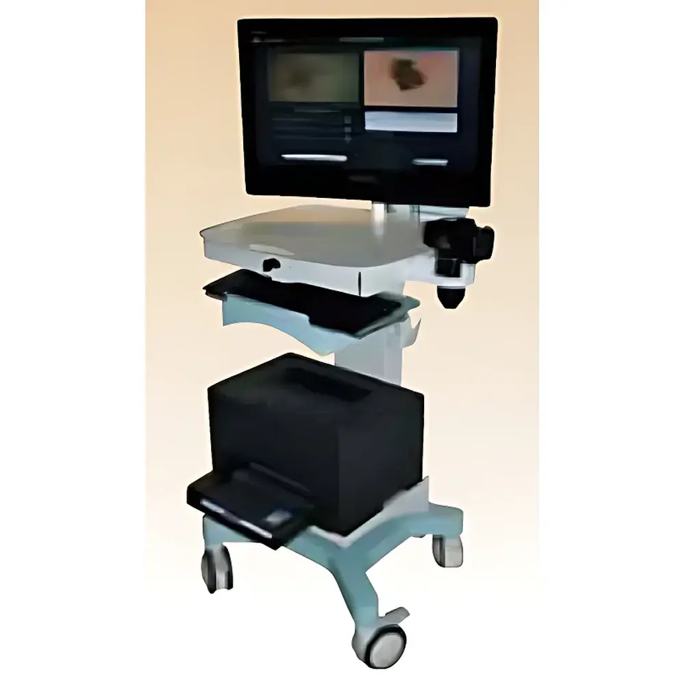
PhotoMAX Trichoscopy & Dermoscopic Imaging Analysis System
| Brand | CK |
|---|---|
| Origin | Austria |
| Manufacturer Status | Authorized Distributor |
| Origin Category | Imported |
| Model | PhotoMAX |
| Pricing | Upon Request |
Overview
The PhotoMAX Trichoscopy & Dermoscopic Imaging Analysis System is a CE-marked, clinical-grade digital imaging platform engineered for standardized, non-invasive assessment of hair and skin morphology. Built upon high-resolution dermoscopic optics and calibrated illumination modules—including polarized and non-polarized ELM (epiluminescence microscopy) modes—the system enables reproducible acquisition of macroscopic, microscopic, and trichoscopic images under controlled lighting and magnification conditions. Its core analytical function in trichoscopy relies on standardized image capture at 20× to 70× magnification, followed by semi-automated morphometric quantification aligned with consensus guidelines from the European Hair Research Society (EHRS) and the International Trichoscopy Society (ITS). Designed for dermatology clinics, trichology centers, and academic research units, PhotoMAX supports longitudinal monitoring of hair density, anagen-to-telogen ratio, hair shaft diameter distribution, and perifollicular inflammation—key endpoints in clinical trials and therapeutic response evaluation.
Key Features
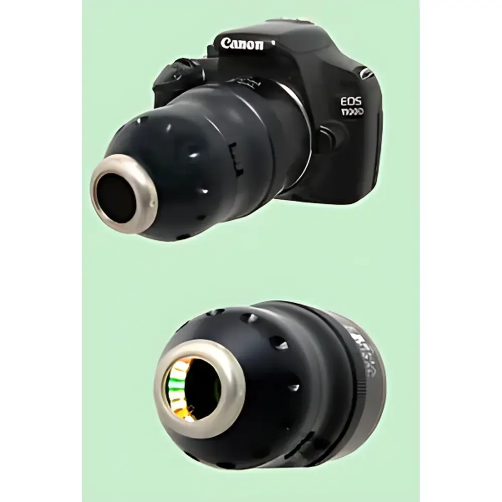
- Multi-modal imaging capability: Simultaneous support for polarized ELM, non-polarized ELM, macro photography (1:1), and contact dermoscopy up to 70× magnification
- Dedicated Trichogram mode with standardized scalp mapping protocol (per ITS-recommended 4–6 cm² sampling zones)
- Integrated calibration reference grid and real-time scale bar overlay for pixel-to-micron conversion
- Adjustable LED illumination intensity and spectral profile optimized for follicular contrast enhancement
- Robust aluminum-alloy housing with ergonomic hand-held probe and fixed stand options for clinic or lab deployment
- Compliance with IEC 62304 (medical device software lifecycle) and EN ISO 13485:2016 quality management requirements
Sample Compatibility & Compliance
The PhotoMAX system is validated for use on Fitzpatrick skin types I–VI and all ethnic hair types (straight, wavy, curly, coily). Scalp imaging requires no topical coupling gel when using the contact dermoscopy mode; non-contact ELM imaging is compatible with dry or lightly prepped (alcohol-wiped) scalp surfaces. All image metadata—including date/time stamp, magnification factor, illumination mode, patient ID, and operator ID—are embedded in DICOM-compliant EXIF headers. The system meets regulatory requirements for Class IIa medical devices under EU MDR 2017/745 and is supplied with a Declaration of Conformity referencing EN 60601-1 (safety) and EN 62304 (software validation). Clinical workflows align with Good Clinical Practice (GCP) documentation standards and support audit-ready data export for regulatory submissions.
Software & Data Management
The proprietary PhotoMAX Analysis Suite (v5.2+) runs on Windows 10/11 64-bit platforms and features a dual-interface architecture: a clinician-facing module for image acquisition and annotation, and a researcher-facing module for batch analysis and statistical reporting. Image storage follows HIPAA- and GDPR-compliant local database architecture (SQLite with AES-256 encryption), with optional integration into hospital PACS via DICOM-SR (Structured Reporting) protocol. Trichoscopic quantification algorithms include automated hair count per mm², anagen/telogen classification based on bulb morphology and pigment distribution (validated against histopathological ground truth in peer-reviewed studies), and cross-sectional hair shaft diameter histogram generation. Audit trails record every image modification, measurement export, and user login event—fully compliant with FDA 21 CFR Part 11 requirements for electronic records and signatures.
Applications
- Longitudinal monitoring of androgenetic alopecia progression and treatment efficacy (e.g., minoxidil, finasteride, low-level laser therapy)
- Objective assessment of telogen effluvium severity and recovery kinetics post-partum or post-illness
- Supporting differential diagnosis between scarring and non-scarring alopecias via perifollicular erythema, scaling, and honeycombing pattern recognition
- Standardized endpoint collection in Phase II–III dermatological clinical trials requiring objective trichoscopic biomarkers
- Teaching and training in trichoscopy interpretation, with built-in reference atlas of >200 annotated clinical cases
FAQ
Is PhotoMAX certified for clinical use in the United States?
PhotoMAX carries CE marking for clinical use in the EU and UK. It is not currently FDA-cleared; U.S. institutions may utilize it for research-only applications under IRB-approved protocols.
Can the system integrate with existing EMR or dermatology practice management software?
Yes—via HL7 v2.x messaging and DICOM-SR export. Custom API integration is available under NDA for enterprise deployments.
What is the recommended maintenance schedule for optical calibration?
CK recommends annual recalibration by an authorized service center using traceable NIST-certified reference targets. In-house verification can be performed weekly using the included calibration slide.
Does the software support multi-user role-based access control?
Yes—administrators can assign roles (e.g., Clinician, Researcher, Technician) with granular permissions for image acquisition, analysis, export, and database administration.
Are raw image files exportable in lossless format?
All acquired images are stored natively in 16-bit TIFF format with embedded metadata; export options include TIFF, PNG, JPEG2000, and DICOM-encapsulated PDF.