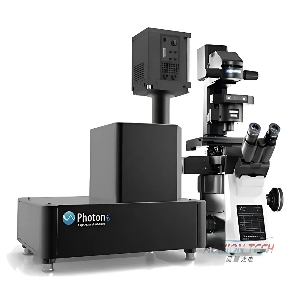
Photon etc IMA Laser-Induced Fluorescence Microscopic Hyperspectral Imaging System
| Brand | Photon etc |
|---|---|
| Origin | Canada |
| Manufacturer Type | Authorized Distributor |
| Product Category | Imported Instrument |
| Model | IMA Laser-Induced Fluorescence Microscopic Hyperspectral Imaging System |
| Spectral Range | 30–6000 cm⁻¹ (Raman) / 400–1700 nm (fluorescence EL/PL) |
| Spectral Resolution | < 0.2 nm (VIS-NIR), < 0.4 nm (SWIR) |
| Spatial Resolution | < 0.02 µm (sub-diffraction limited, objective-dependent) |
| Spectral Reproducibility | < 0.1 nm RMS over 8-hour operation |
| Excitation Wavelengths | 447, 532, 635, 808, 980 nm (customizable) |
| Detector Options | Back-illuminated sCMOS (VIS-NIR), InGaAs array (SWIR), EMCCD (low-light PL) |
| Imaging Modality | Widefield non-scanning hyperspectral acquisition using Volume Bragg Tunable Filter (BTF) technology |
| Software Platform | PHySpec™ v5.2 with PCA, MCR, spectral unmixing, time-resolved spectral video export, and FDA 21 CFR Part 11–compliant audit trail |
Overview
The Photon etc IMA Laser-Induced Fluorescence Microscopic Hyperspectral Imaging System is an advanced widefield platform engineered for simultaneous spatial and spectral characterization of luminescent materials at sub-micron resolution. Unlike conventional point-scanning confocal or Raman microscopes, the IMA employs a patented Volume Bragg Grating (VBG)-based tunable filter architecture to acquire full hyperspectral cubes—spanning 400–1700 nm (with optional Raman extension to 6000 cm⁻¹)—in a single exposure per wavelength step. This non-scanning, snapshot-based acquisition eliminates mechanical raster delays and photobleaching risks associated with sequential scanning, enabling quantitative electroluminescence (EL) and photoluminescence (PL) mapping of delicate biological specimens, nanomaterials, and optoelectronic devices without sample damage. The system integrates seamlessly with inverted or upright research-grade microscopes and supports dual-modal operation: high-throughput fluorescence hyperspectral imaging in the visible–shortwave infrared (VIS-SWIR) range, and high-fidelity Raman spectroscopic imaging via external laser coupling and notch filtering.
Key Features
- Widefield hyperspectral acquisition: Captures full 2D spatial + 1D spectral datacubes (e.g., 1024 × 1024 × 256) without point-by-point scanning—reducing acquisition time by >100× compared to confocal equivalents.
- Volume Bragg Tunable Filter (BTF) core: Enables precise, repeatable spectral tuning (OD6), eliminating moving parts and thermal hysteresis common in liquid crystal or acousto-optic tunable filters.
- Sub-diffraction spatial resolution: Achieves <20 nm lateral resolution under 100× oil immersion (λ = 532 nm, NA = 1.49), validated via calibrated nanoparticle test targets and NIST-traceable line-pair standards.
- Dual-detector compatibility: Supports scientific-grade back-illuminated sCMOS for VIS-NIR (400–1000 nm) and thermoelectrically cooled InGaAs arrays for SWIR (900–1700 nm), both synchronized to BTF stepping with <10 µs timing jitter.
- Multi-wavelength excitation flexibility: Integrated laser combiner accepts up to five solid-state lasers (447, 532, 635, 808, 980 nm), each independently modulated for time-gated or ratiometric PL/EL studies.
- Quantitative radiometric calibration: Factory-calibrated absolute intensity response traceable to NIST standards; includes two-step protocol: point-source laser calibration followed by integrating sphere–based spatial-spectral uniformity correction.
Sample Compatibility & Compliance
The IMA system accommodates diverse sample formats—including live cells on glass-bottom dishes, tissue cryosections, thin-film solar cell wafers, and nanomaterial-coated substrates—without requiring vacuum or conductive coating. Its non-contact, low-fluence widefield illumination (typically <10⁷ W/m²) preserves viability in live-cell assays, as demonstrated in longitudinal tracking of 60 nm gold nanoparticle uptake in MDA-MB-231 breast cancer lines over 72 hours. For regulated environments, the system complies with ISO/IEC 17025 analytical instrument qualification requirements and supports GLP/GMP workflows through PHySpec™’s embedded audit trail, electronic signatures, and 21 CFR Part 11–compliant user access controls. All optical components meet RoHS and REACH directives; laser safety conforms to IEC 60825-1:2014 Class 3B/4 interlock protocols.
Software & Data Management
PHySpec™ v5.2 serves as the unified acquisition, processing, and reporting engine. It provides real-time spectral cube streaming to RAM or SSD (up to 1.2 GB/s sustained write speed), GPU-accelerated multivariate analysis—including principal component analysis (PCA), multivariate curve resolution (MCR), and constrained non-negative matrix factorization (CNMF)—and batch-processed spectral unmixing for multiplexed SWCNT chirality identification. Export options include HDF5 (with metadata schema compliant with FAIR principles), TIFF stacks with embedded EXIF spectral tags, and CSV-formatted spectra for integration into MATLAB, Python (scikit-learn, HyperSpy), or commercial chemometrics packages. Raw data integrity is ensured via SHA-256 checksum logging; all processing steps are recorded in immutable audit trails meeting FDA and EMA regulatory expectations for analytical method validation.
Applications
- Nanoparticle biodistribution & speciation: Discrimination of single-wall carbon nanotube (SWCNT) chiralities (n,m) via narrowband NIR emission (900–1700 nm), enabling multiplexed detection of ≥17 distinct species in live murine macrophages with subcellular localization accuracy.
- Photovoltaic device metrology: Quantitative EL/PL cartography of multicrystalline CIS and GaAs solar cells; extraction of spatially resolved open-circuit voltage (VOC) maps, carrier diffusion length gradients, and defect cluster identification—validated against calibrated reference cells per IEC 60904-8.
- Biological dark-field hyperspectral imaging: High-contrast label-free detection of plasmonic nanoparticles in cellular cytoplasm using scattering spectral fingerprints; PCA-driven segmentation separates aggregated vs. monodisperse 60 nm AuNPs based on peak redshift magnitude and bandwidth broadening.
- In vivo spectral sensing: Longitudinal monitoring of SWCNT-based biosensors in murine tumor models, leveraging second biological window (NIR-II) advantages: reduced autofluorescence, deeper penetration (>3 mm), and minimal scattering-induced spectral distortion.
FAQ
Does the IMA support true confocal operation?
No—the IMA is a widefield hyperspectral imager. It does not incorporate pinhole-based optical sectioning. However, its sub-micron spatial resolution and axial discrimination via high-NA objectives enable effective optical sectioning in thick samples when combined with deconvolution algorithms in PHySpec™.
Can Raman and fluorescence modes be used simultaneously?
Not concurrently. The system requires optical reconfiguration: fluorescence mode uses the integrated BTF + sCMOS/InGaAs path; Raman mode requires external 532/785 nm lasers, holographic notch filters, and a dedicated spectrometer-coupled CCD. Switching takes <15 minutes and is documented in the IQ/OQ protocol.
Is spectral calibration traceable to national standards?
Yes—each instrument ships with NIST-traceable calibration certificates for wavelength (Hg/Ne lamp lines), intensity (integrating sphere + calibrated photodiode), and spatial scale (NIST SRM 2051 grating standard). Annual recalibration services are available through Photon etc’s ISO/IEC 17025-accredited lab.
What is the minimum detectable PL quantum yield?
Under optimal conditions (100×, 532 nm excitation, EMCCD, 2 s integration), the system achieves a limit of detection of ~10⁻⁴ photons/pixel/s for narrowband emitters (FWHM < 5 nm), corresponding to quantum yields ≥10⁻⁵ for diffraction-limited spots.
Related Products


")