PSI FKM Multispectral Fluorescence Kinetic Microscope
| Brand | PSI |
|---|---|
| Origin | Czech Republic |
| Model | FKM |
| Price Range | USD 68,000 – 136,000 |
| Excitation Light Sources | IR, Red, Orange, Blue, Green, White, UV, Far-Red |
| Spectral Range (SM9000 Spectrometer) | 200–980 nm |
| Wavelength Accuracy | <0.5 nm |
| CCD Resolution | 1360 × 1024 pixels |
| Frame Rate | up to 20 fps at full resolution |
| A/D Depth | 16-bit (65,536 gray levels) |
| Temperature Control Range | 5–70 °C (±0.1 °C) |
| Objective Lenses | 10×, 20×, 40×, 63×, 100× fluorescence-grade |
| Filter Wheel | 6-position, configurable for Chl a, GFP/SYTOX, DAPI/CTC, etc. |
Overview
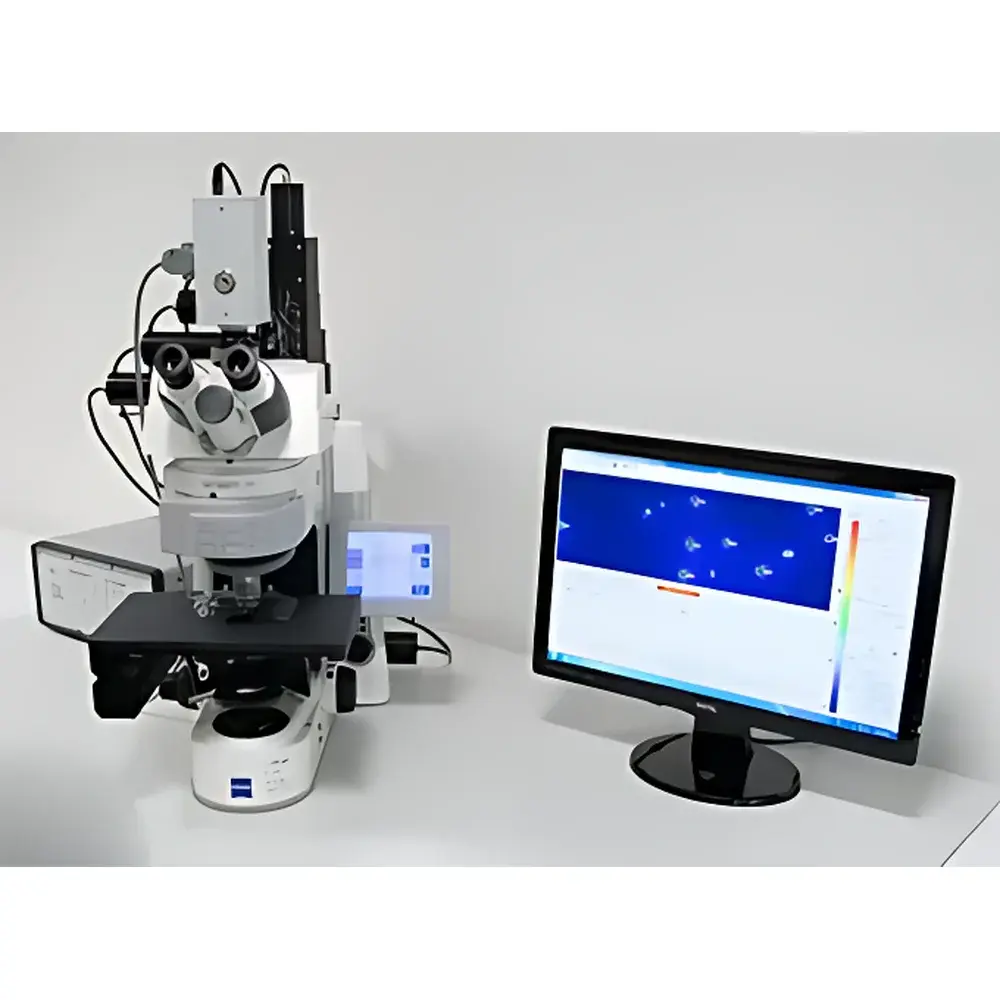
The PSI FKM Multispectral Fluorescence Kinetic Microscope is a research-grade, integrated platform engineered for quantitative, subcellular-level analysis of photosynthetic function and multicolor fluorescence dynamics in living plant and algal systems. Built upon the foundational principles of pulse-amplitude-modulated (PAM) chlorophyll fluorescence imaging and extended to microscopic spatial resolution, the FKM combines high-temporal-resolution kinetic microscopy with multispectral detection and spectral deconvolution capabilities. Its core measurement methodology relies on controlled photochemical excitation—via programmable, spectrally defined light sources—and time-resolved detection of emitted fluorescence across multiple emission bands, enabling simultaneous quantification of photosystem II (PSII) quantum yield, non-photochemical quenching (NPQ), electron transport rate (ETR), and OJIP transient kinetics. The system further extends beyond chlorophyll a fluorescence by supporting exogenous fluorophore imaging (e.g., GFP, DAPI, DiBAC₄, SYTOX) and intrinsic pigment spectroscopy (e.g., phycobilins, carotenoids), making it uniquely suited for correlative functional phenotyping at the organelle, cell, and tissue level.
Key Features
- Integrated high-magnification fluorescence microscope (Zeiss Axio Imager M2 or configurable alternatives) with automated 7-position objective turret and precision 6-position filter wheel for sequential multi-channel acquisition.
- Dual-detection architecture: high-sensitivity, 16-bit TOMI-2 CCD camera (1360 × 1024 px, 20 fps max) for kinetic imaging; SM9000 fiber-coupled spectrometer (200–980 nm, <0.5 nm absolute accuracy) for spectral fingerprinting of individual fluorophores and photosynthetic complexes.
- Modular excitation source array including narrowband LEDs and broadband sources covering UV (365 nm), violet, blue (450 nm), green (520 nm), orange (590 nm), red (630 nm), far-red (735 nm), and near-infrared (780 nm), enabling selective activation of PSII, PSI, cryptochromes, phytochromes, and exogenous probes.
- Programmable temperature control module (5–70 °C, ±0.1 °C stability) for reproducible physiological assays and controlled abiotic stress imposition (heat, cold, thermal ramping).
- Full-featured FluorCam software suite with protocol-driven automation, ROI-based parametric mapping (>1000 regions per image), dual signal-processing modes (“signal-average-then-calculate” vs. “calculate-then-average”), and timestamped, audit-ready data export compliant with GLP documentation standards.
Sample Compatibility & Compliance
The FKM supports live, non-invasive measurements across a broad biological scope: intact leaf epidermis, enzymatically isolated mesophyll protoplasts, purified chloroplasts and thylakoid membranes, unicellular and colonial microalgae (e.g., Chlamydomonas, Synechocystis, Dunaliella), cyanobacterial heterocysts, and symbiotic systems (e.g., coral zooxanthellae, lichen photobionts). All optical and thermal modules are calibrated traceably to NIST standards. Software workflows support 21 CFR Part 11-compliant electronic signatures, user access control, and immutable audit trails—essential for regulated preclinical or industrial R&D environments. Hardware and firmware comply with IEC 61000-6-3 (EMC) and IEC 61010-1 (safety) requirements for laboratory instrumentation.
Software & Data Management
FluorCam v7.x provides a unified interface for experimental design, acquisition, and advanced post-processing. Users define custom protocols using a structured scripting language or guided wizards—specifying light intensity (μmol photons·m⁻²·s⁻¹), duration, sequence order, dark adaptation intervals, and trigger logic. Acquired datasets include synchronized time-series images, spectral scans per pixel (hyperspectral mode), and kinetic parameter maps (e.g., ΦPSII, NPQ, qP, PIABS). Data are stored in vendor-neutral HDF5 format with embedded metadata (instrument configuration, environmental conditions, operator ID). Batch analysis supports statistical comparison across genotypes, treatments, or time points, with output formats including Excel (.xlsx), TIFF stacks, AVI video, and interactive HTML reports. Optional integration with MATLAB and Python via documented APIs enables custom algorithm development and machine-learning–based phenotype classification.
Applications
- Subcellular mapping of PSII/PSI spatial heterogeneity and repair dynamics under fluctuating light.
- High-throughput screening of photosynthetic mutants and transgenic lines for altered NPQ capacity or electron transport efficiency.
- Quantitative assessment of biotic (pathogen, symbiont) and abiotic (drought, salinity, heavy metal, temperature) stress responses at the chloroplast level.
- Correlative imaging of chlorophyll fluorescence with nuclear DNA integrity (DAPI), membrane potential (DiBAC₄), or viability (SYTOX) in single cells.
- Resolving spectral contributions of phycocyanin, allophycocyanin, and phycoerythrin in cyanobacteria and red algae under nutrient limitation.
- In vivo kinetic modeling of QA⁻ reoxidation and intersystem electron pool size using millisecond-scale flash sequences.
- Time-lapse phenotyping of microalgal cultures under continuous perfusion (via optional peristaltic pump), enabling long-term photophysiological monitoring without manual intervention.
FAQ
What distinguishes the FKM from conventional macro-scale FluorCam systems?
The FKM integrates diffraction-limited optical magnification with microsecond-precision light control and spectral deconvolution—enabling fluorescence quantification at the single-organelle level, which is not resolvable in wide-field leaf-level imaging.
Can the FKM perform simultaneous dual-emission detection (e.g., Chl a + GFP)?
Yes—using the 6-position filter wheel and time-gated acquisition, users can interleave excitation/emission channels within a single kinetic protocol without cross-talk.
Is spectral calibration required before each experiment?
No—the SM9000 spectrometer includes internal wavelength reference and thermal stabilization; factory calibration remains valid for ≥12 months under standard lab conditions.
Does the system support third-party microscope objectives?
Only Zeiss-certified fluorescence objectives are validated for optimal transmission, chromatic correction, and working distance compatibility with the integrated light path and filter wheel geometry.
How is data integrity ensured during unattended overnight measurements?
All acquisitions are logged with hardware timestamps, checksum-verified file writes, and automatic error recovery; failed cycles trigger alerts and resume from last valid checkpoint.


