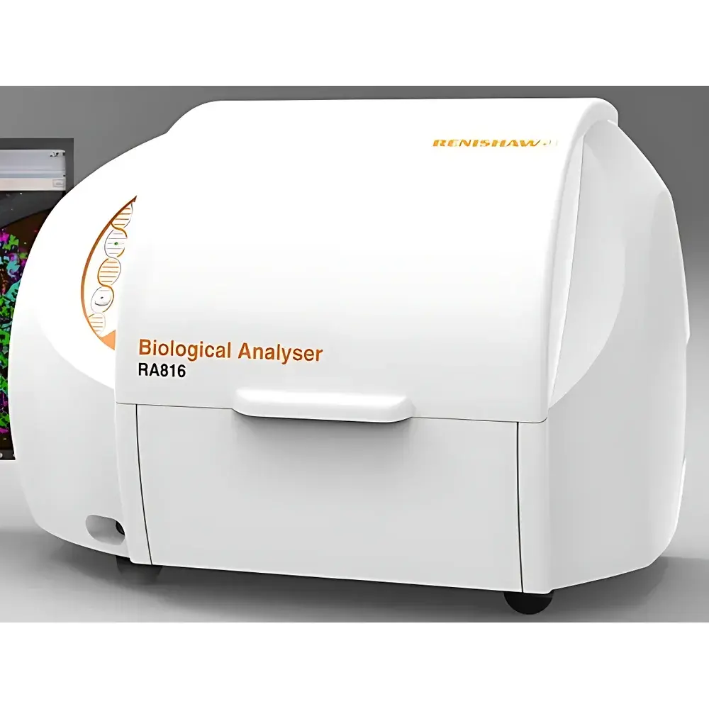
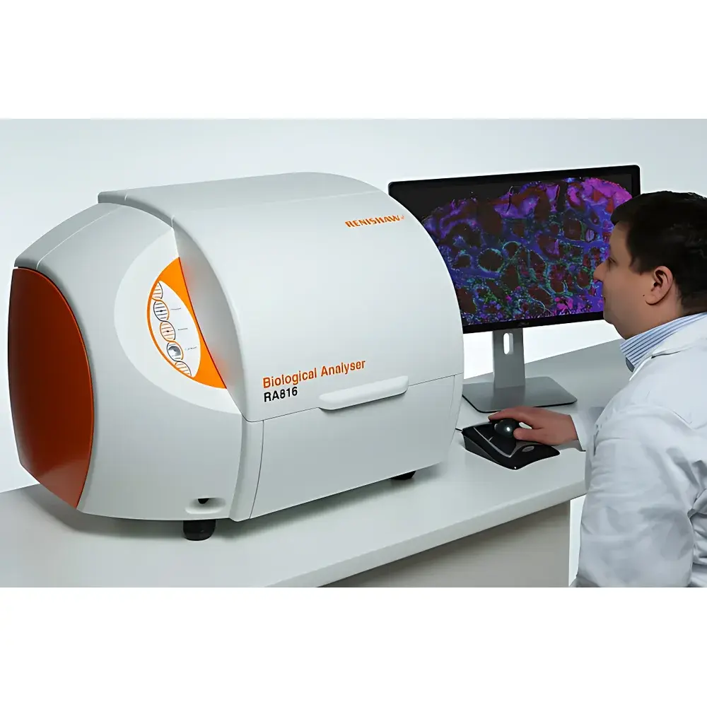
Renishaw RA816 Biological Analyser
| Brand | Renishaw |
|---|---|
| Origin | United Kingdom |
| Model | RA816 |
| Instrument Type | Confocal Micro-Raman Spectrometer |
| Spectral Range | 100–3500 cm⁻¹ |
| Spectral Resolution | 2 cm⁻¹ |
| Spatial Resolution | ~500 nm |
| Minimum Wavenumber | 100 cm⁻¹ |
| Spectral Reproducibility | ≤ ±0.1 cm⁻¹ |
Overview
The Renishaw RA816 Biological Analyser is a benchtop confocal micro-Raman spectrometer engineered for high-fidelity biochemical characterisation of unmodified biological specimens. Operating on the principle of inelastic light scattering—where monochromatic laser excitation induces vibrational mode shifts in molecular bonds—the system delivers label-free, non-destructive spectral fingerprints with subcellular spatial registration. Unlike conventional widefield Raman systems, the RA816 integrates a true confocal optical architecture with motorised XYZ stage control and automated focus tracking, enabling depth-resolved spectral acquisition from heterogeneous tissue volumes. Its design prioritises robustness in regulated laboratory environments, supporting reproducible data generation across multi-user academic, clinical research, and translational biopharma settings.
Key Features
- Confocal optical design with adjustable pinhole aperture for axial sectioning and rejection of out-of-focus fluorescence background
- Integrated 785 nm diode laser (optional 532 nm or 633 nm modules available) with thermoelectric cooling and power stability < ±0.5% over 8 hours
- High-throughput CCD detector cooled to –70 °C for low-noise spectral acquisition (typical integration times: 0.1–30 s per pixel)
- Automated XYZ stage with 100 nm step resolution and closed-loop feedback for precise raster mapping
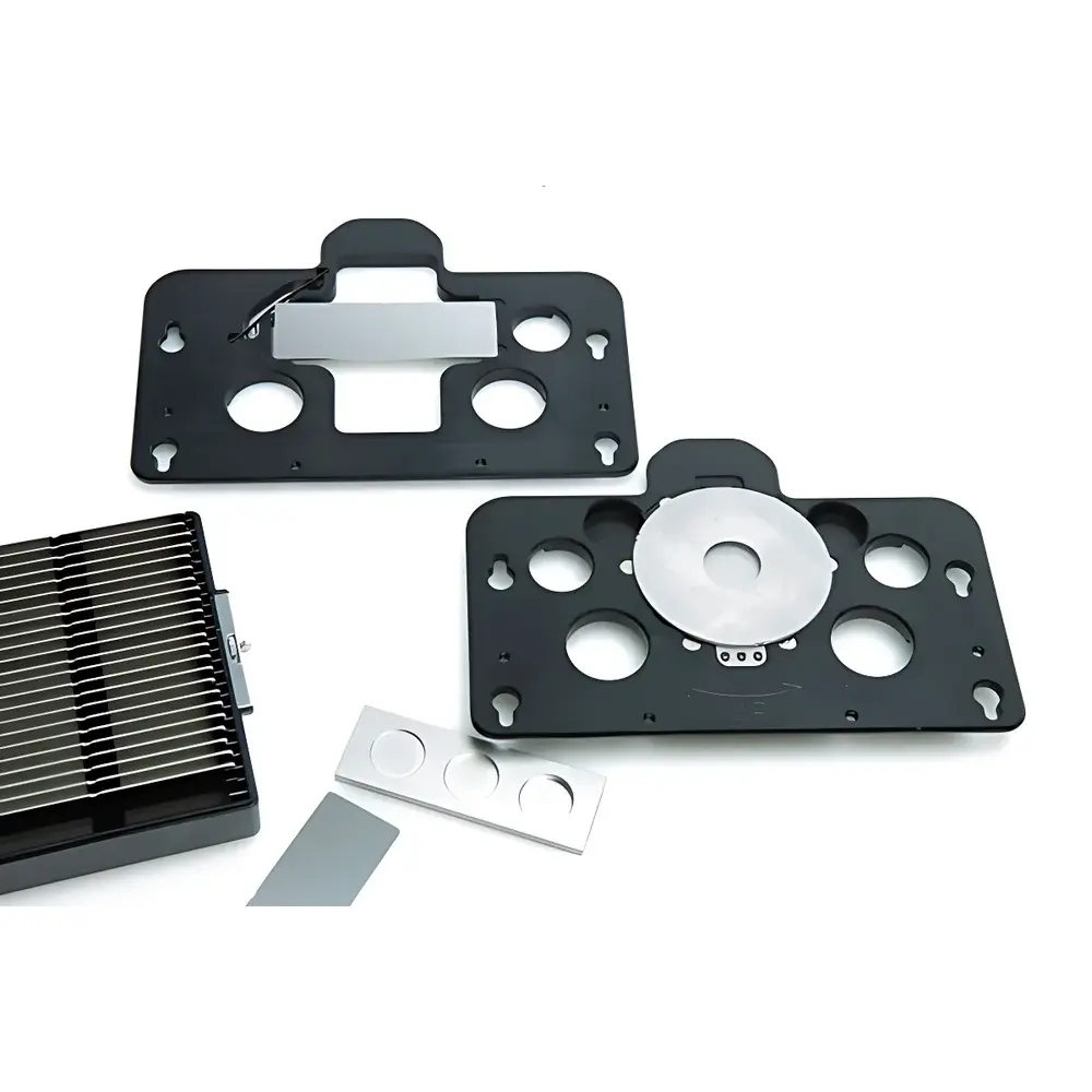
- Dedicated biological sample interface: motorised objective turret (×20, ×50, ×100 dry/water-immersion options), environmental chamber compatibility (temperature/humidity control), and integrated brightfield imaging for co-registered morphological context
- Pre-aligned optics and factory-calibrated wavelength scale (NIST-traceable polystyrene reference) ensure day-one operational readiness without user alignment
Sample Compatibility & Compliance
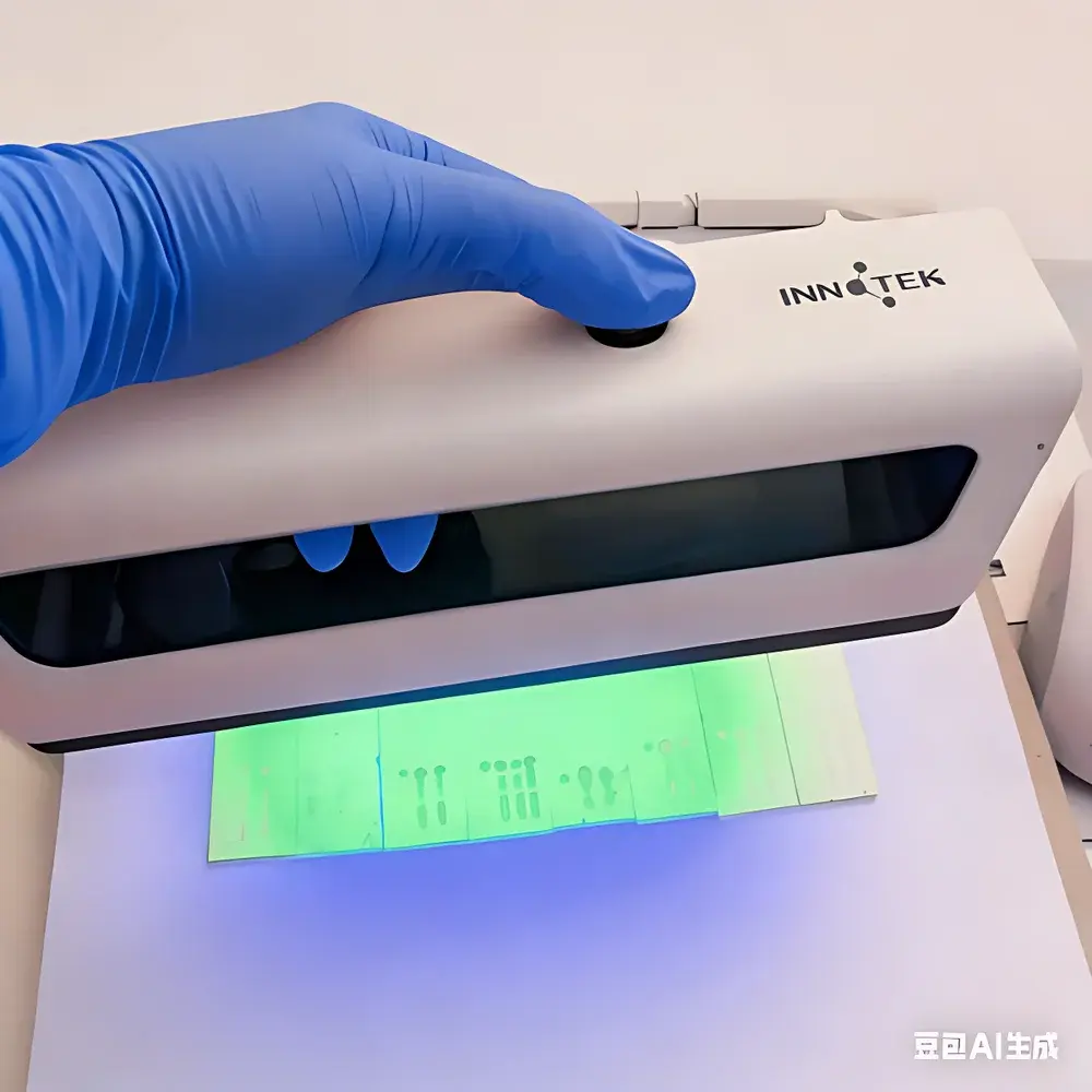
The RA816 is validated for direct analysis of native, hydrated, and fixed biological matrices—including formalin-fixed paraffin-embedded (FFPE) tissue sections, fresh-frozen cryosections, cell monolayers, biofluids (serum, saliva, CSF), and microbial colonies—without chemical labelling or metal enhancement. Sample mounting follows standard histopathology protocols (glass slides, CaF₂ windows, or quartz substrates). The system complies with ISO/IEC 17025:2017 requirements for analytical instrument validation and supports audit-ready documentation per GLP and GMP frameworks. Spectral data integrity adheres to FDA 21 CFR Part 11 principles via electronic signatures, audit trails, and role-based access control within the bundled WiRE™ software platform.
Software & Data Management
Controlled by Renishaw’s WiRE 5.x software suite, the RA816 provides end-to-end workflow management—from real-time spectral preview and auto-exposure optimisation to multivariate chemometric analysis (PCA, cluster analysis, spectral unmixing). Raw spectra are stored in vendor-neutral .spc and HDF5 formats; metadata (laser power, objective, integration time, stage coordinates) is embedded per spectrum. Batch processing supports large-area hyperspectral maps (>10⁶ pixels), with GPU-accelerated denoising and baseline correction algorithms. Export modules enable seamless integration with third-party platforms including MATLAB, Python (via PySpectra), and commercial pathology informatics systems (e.g., 3DHISTECH, Visiopharm).
Applications
- Label-free histopathology: discrimination of tumour margins, necrotic vs. viable regions, and stromal composition in FFPE and frozen sections
- Single-cell phenotyping: metabolic profiling of immune cells, stem cells, and circulating tumour cells in suspension or adherent culture
- Drug distribution mapping: spatial quantification of small-molecule therapeutics and metabolites within tissue microenvironments
- Biofluid biomarker discovery: detection of low-abundance protein conformational changes or lipid peroxidation products in serum or cerebrospinal fluid
- Microbial identification: strain-level classification of bacteria and fungi based on conserved ribosomal and membrane lipid signatures
- Bioprocess monitoring: real-time assessment of extracellular matrix deposition and cellular differentiation in 3D bioreactors and organoids
FAQ
Is the RA816 suitable for live-cell Raman imaging?
Yes—when paired with environmental control accessories (stage-top incubator, CO₂ regulation, and low-laser-power protocols), the RA816 enables time-lapse Raman mapping of living cells with minimal phototoxicity.
Can it be integrated into existing laboratory information management systems (LIMS)?
Yes—WiRE supports HL7 and ASTM E1467-compliant data export, and custom API hooks allow bidirectional communication with enterprise LIMS and ELN platforms.
What calibration standards are supplied with the instrument?
The system ships with NIST-traceable polystyrene and silicon wafers for daily wavelength and intensity calibration, plus a certified reference material (CRM) set for biological matrix validation (e.g., bovine serum albumin, collagen I, and DNA standards).
Does the RA816 meet regulatory requirements for clinical assay development?
While not an IVD device, the RA816 satisfies key analytical validation criteria outlined in CLSI EP17-A2 and ISO 20943 for method development in clinical research laboratories pursuing CE-IVDR or FDA pre-submission pathways.