Retinal Vessel Analyzer RVA-Static VesselMap System
No specifications available.
Overview
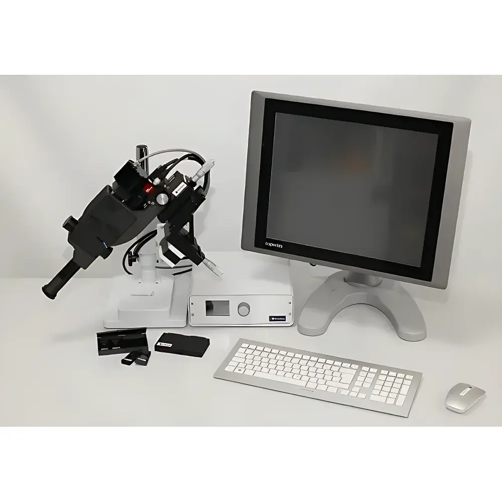
The Retinal Vessel Analyzer RVA-Static VesselMap System is a clinical-grade ophthalmic imaging and quantitative analysis platform engineered for standardized, semi-automatic assessment of retinal microvasculature morphology. It operates on the principle of high-resolution fundus photography combined with validated image segmentation algorithms to extract geometric parameters from retinal vessel trees. The system implements the standardized protocols established by the Wisconsin Epidemiologic Study of Diabetic Retinopathy (WESDR) and the Atherosclerosis Risk in Communities (ARIC) study, ensuring inter-study comparability and longitudinal reproducibility. Designed for use in academic ophthalmology departments, clinical research units, and epidemiological cohort studies, the RVA-Static VesselMap System delivers objective, operator-independent quantification of structural vascular biomarkers associated with systemic hypertension, diabetes mellitus, cerebrovascular disease, and early-stage microvascular dysfunction.
Key Features
- Semi-automated vessel detection and centerline tracing using adaptive thresholding and morphological filtering optimized for 45°–60° non-mydriatic or mydriatic fundus images
- Standardized calculation of the Central Retinal Arterial Equivalent (CRAE) and Central Retinal Venous Equivalent (CRVE) based on the Parr-Hubbard formula, incorporating vessel width, branching geometry, and spatial weighting across the optic disc-centered zone (0.5–1.0 disc diameters)
- Automated derivation of the Arterio-Venous Ratio (AVR = CRAE / CRVE), a validated surrogate marker of systemic vascular resistance and endothelial health
- Integrated quality control module that flags low-contrast images, motion artifacts, or incomplete optic disc capture prior to analysis
- Modular software architecture compliant with DICOM-SOP Class 1.2.840.10008.5.1.4.1.1.77.1.5.4 (Ophthalmic Photography Image Storage)
- Export functionality supporting CSV, XML, and HL7 CDA v2.0 formats for integration into electronic health record (EHR) systems and clinical trial databases
Sample Compatibility & Compliance
The system accepts standard color or monochrome fundus photographs acquired with commercially available desktop or portable fundus cameras (e.g., Topcon TRC-NW series, Zeiss FF 450+, Canon CR-2 Plus). Input images must meet minimum resolution requirements (≥1600 × 1200 pixels) and include visible optic disc margins and at least three major arteriolar and venular branches within the measurement zone. The analytical pipeline adheres to ISO 13485:2016 medical device quality management standards and supports audit-ready documentation per FDA 21 CFR Part 11 requirements, including user authentication logs, version-controlled algorithm parameters, and immutable analysis timestamps. All output metrics comply with the reporting conventions defined in the STROBE-Ophth extension for observational ophthalmic studies.
Software & Data Management
VesselMap Analysis Suite v4.x provides a secure, role-based desktop interface with configurable user permissions (administrator, clinician, researcher, data manager). Raw images are stored in AES-256 encrypted local repositories; analysis metadata—including pixel calibration coefficients, vessel selection masks, and confidence scores—is embedded in XMP sidecar files. The software supports batch processing of longitudinal image series and generates PDF reports conforming to ICH E6(R3) Good Clinical Practice guidelines. Audit trails capture every parameter modification, export event, or user login, enabling full traceability during regulatory inspections. Integration with REDCap and OpenClinica is supported via RESTful API endpoints with OAuth 2.0 authentication.
Applications
- Epidemiological monitoring of microvascular aging in population-based cohorts (e.g., UK Biobank, Rotterdam Study)
- Endpoint quantification in interventional trials evaluating antihypertensive or anti-angiogenic therapies
- Early detection of cerebral small vessel disease through retinal-vascular correlation modeling
- Validation of AI-based retinal biomarker algorithms against gold-standard manual grading
- Training platform for standardized retinal vessel measurement in ophthalmology residency programs
- Longitudinal tracking of diabetic retinopathy progression independent of conventional grading scales
FAQ
What image acquisition protocol is required for optimal CRAE/CRVE calculation?
A centered 45° or 50° color fundus photograph with clear optic disc margins, minimal lens flare, and adequate illumination uniformity across the posterior pole is required. Mydriasis is recommended but not mandatory if image quality meets SNR >25 dB.
Does the system support stereo pair analysis or dynamic (flicker) vessel response measurements?
No—this is a static vessel analysis platform. Dynamic functional assessments (e.g., Flicker Light-Induced Retinal Vasodilation) require the companion RVA-Dynamic module.
Can CRAE and CRVE values be exported directly to SPSS or SAS for statistical modeling?
Yes—CSV exports include subject ID, visit date, CRAE (µm), CRVE (µm), AVR, measurement confidence index, and image QA status, formatted for direct ingestion into common statistical packages.
Related Products