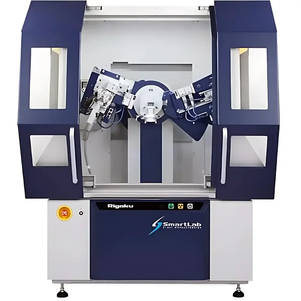
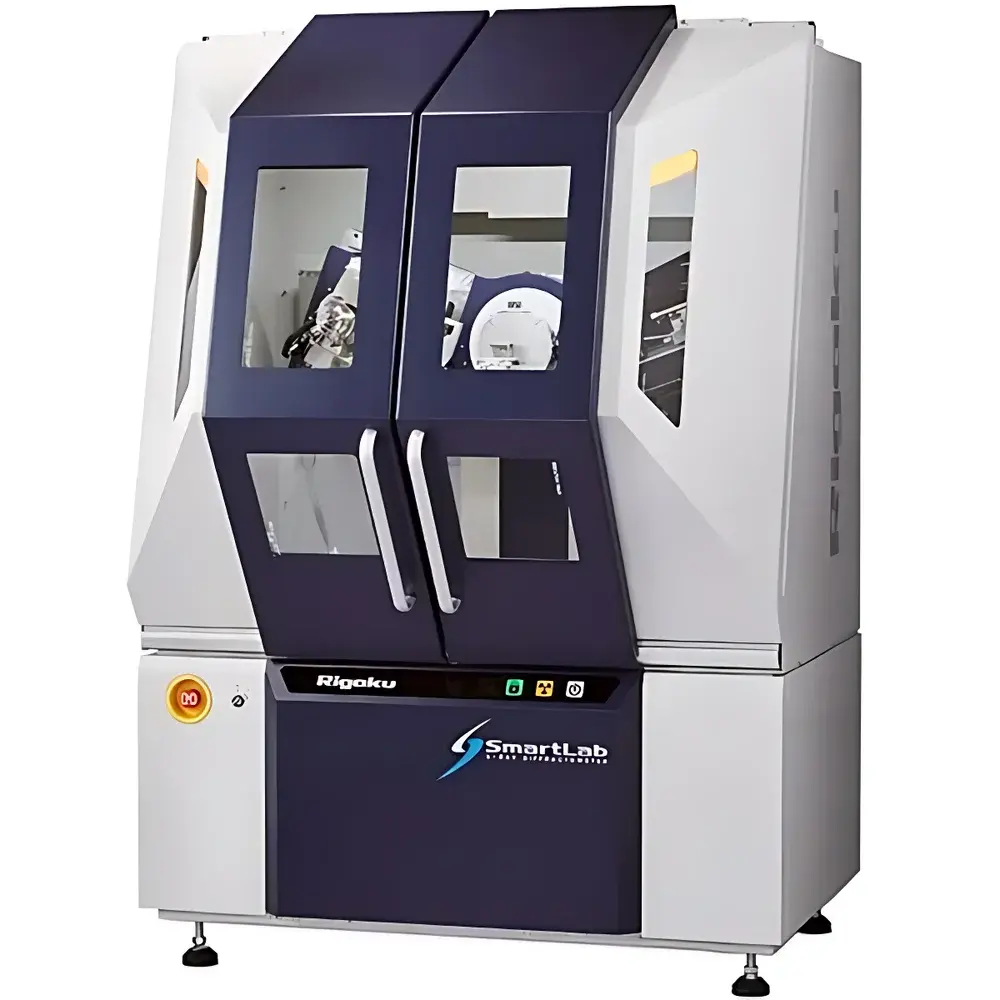
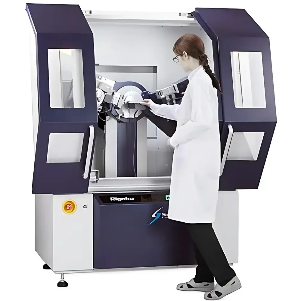
Rigaku SmartLab Automated Powder X-ray Diffractometer
| Brand | Rigaku |
|---|---|
| Origin | Japan |
| Instrument Type | Powder X-ray Diffractometer |
| Model | SmartLab |
| X-ray Source | 3 kW or 9 kW sealed-tube generator |
| Anode Materials | Cu, Cr, Fe, Co, Ni, Mo, Ag, Au (selectable) |
| Goniometer Radius | 300 mm |
| Detector | HyPix-3000 2D pixel array detector |
| Optics | CBO-Auto (automated beam optics switching) and CBO-μ (high-resolution micro-beam optics) |
| Primary Source | PhotonMax high-brilliance X-ray tube |
| Software | SmartLab Studio II (with in-situ analysis modules) |
Overview
The Rigaku SmartLab Automated Powder X-ray Diffractometer is a high-precision, fully automated benchtop system engineered for rigorous phase identification, quantitative phase analysis (QPA), crystallite size and microstrain determination, residual stress measurement, and thin-film characterization. It operates on the fundamental principle of Bragg’s Law (nλ = 2d sinθ), utilizing monochromatic X-ray radiation to probe the periodic lattice structure of crystalline materials. The instrument employs θ–2θ scanning geometry with a fixed divergence slit and variable receiving slits, enabling high angular resolution and exceptional peak fidelity across the full 2θ range (typically −5° to 145°). Its modular architecture integrates a high-stability goniometer with 300 mm radius, ensuring mechanical reproducibility better than ±0.0005° in 2θ — critical for long-term calibration stability and inter-laboratory data comparability.
Key Features
- PhotonMax X-ray source: High-brilliance sealed-tube generator available in 3 kW (standard) or 9 kW (high-power) configurations, delivering superior photon flux without requiring water cooling in standard operation.
- HyPix-3000 2D pixel array detector: Zero-noise, single-photon counting detector with 300 × 300 pixels, energy discrimination capability, and real-time data collection at frame rates up to 100 Hz — enabling rapid acquisition of Debye–Scherrer rings and simultaneous multi-angle analysis.
- CBO (Cross-Beam Optics) platform: Fully motorized, software-controlled optical path selection between CBO-Auto (for high-throughput routine analysis) and CBO-μ (micro-beam mode with sub-100 µm spot size and optimized for thin films, small samples, or heterogeneous materials).
- SmartLab Studio II software suite: Integrated environment supporting Rietveld refinement (via PDXL), whole-pattern fitting, PDF analysis, in-situ stage control (temperature, humidity, gas atmosphere), and automated method sequencing with audit-trail logging compliant with GLP and 21 CFR Part 11 requirements.
- Robust mechanical architecture: Precision-ground granite base, temperature-compensated goniometer bearings, and vibration-damped enclosure ensure long-term positional stability under varying ambient conditions.
Sample Compatibility & Compliance
The SmartLab accommodates standard powder specimens (≥10 mg, particle size < 10 µm), bulk solids, thin films (on Si wafers or glass substrates), fibers, and polycrystalline metals. Optional accessories include HTK-1200N high-temperature stage (RT–1200 °C), TTK-450 cryo-stage (−180 °C to +450 °C), and environmental cells for in-situ gas/liquid reactions. All hardware and software modules conform to ISO 17025:2017 general requirements for testing laboratories. Data acquisition and processing workflows support traceable calibration per ASTM E975 (Standard Practice for X-ray Diffraction Crystallographic Analysis), ISO 13126 (XRD for pharmaceuticals), and USP (Powder X-ray Diffraction). Audit trails, electronic signatures, and user-access controls are implemented in accordance with FDA 21 CFR Part 11 for regulated environments.
Software & Data Management
SmartLab Studio II provides a unified interface for instrument control, real-time visualization, and advanced data reduction. It features automated background subtraction, peak search with adaptive thresholding, and seamless integration with ICDD PDF-4+ database (2023 edition). Batch processing supports parallel refinement of multiple datasets using identical structural models. Raw 2D detector frames are stored in vendor-neutral HDF5 format; processed patterns export to CIF, XYE, and CSV formats. Network deployment enables centralized license management and remote monitoring via secure HTTPS. All user actions — including parameter changes, calibration events, and report generation — are timestamped and logged with operator ID for full traceability.
Applications
- Pharmaceutical solid-state characterization: polymorph screening, hydrate/anhydrate differentiation, amorphous content quantification.
- Materials science research: crystal structure solution from powder data, lattice parameter refinement, texture analysis via pole figure mapping.
- Geology and mining: mineral phase identification in complex ores, clay mineral quantification, thermal alteration studies.
- Quality control in ceramics and battery materials: cathode/anode crystallinity assessment, degradation monitoring after cycling.
- Nanomaterials analysis: Scherrer-based crystallite size distribution, Williamson–Hall microstrain decomposition, defect density estimation.
FAQ
What anode materials are supported, and how is wavelength selection managed?
The SmartLab accepts sealed-tube anodes in Cu, Cr, Fe, Co, Ni, Mo, Ag, and Au. Wavelength selection is achieved by changing the tube assembly; no monochromator swapping is required. Cu Kα (1.5418 Å) remains the default for most applications due to optimal balance of penetration depth and scattering power.
Is the system compatible with third-party in-situ stages?
Yes — the SmartLab Studio II API supports custom stage integration via RS-232, USB, or Ethernet protocols. Rigaku-certified stages (e.g., HTK-1200N, TTK-450) are pre-configured with thermal compensation and synchronized data logging.
How does the CBO-μ micro-beam configuration achieve spatial resolution?
CBO-μ utilizes nested Göbel mirrors and a motorized pinhole collimator to generate a focused beam ≤80 µm FWHM at the sample position, enabling diffraction mapping with step resolution down to 10 µm without scanning the detector.
Can raw 2D detector images be exported for external processing?
Yes — all HyPix-3000 frames are saved in HDF5 format with embedded metadata (exposure time, motor positions, voltage/current), supporting import into GSAS-II, TOPAS, or custom Python/Matlab workflows.
What level of software validation support is provided for regulated labs?
Rigaku delivers IQ/OQ documentation packages, installation qualification checklists, and a 21 CFR Part 11 readiness guide. Full validation services, including PQ protocol development and UAT support, are available through certified field application specialists.
Related Products