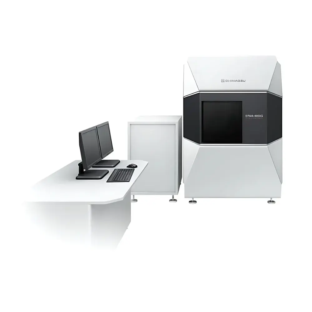
Shimadzu EPMA-8050G Field-Emission Electron Probe Microanalyzer
| Brand | Shimadzu |
|---|---|
| Origin | Japan |
| Manufacturer | Shimadzu Corporation |
| Type | Imported Instrument |
| Model | EPMA-8050G |
| Pricing | Available Upon Request |
Overview
The Shimadzu EPMA-8050G Field-Emission Electron Probe Microanalyzer represents a paradigm shift in quantitative microchemical analysis at submicron spatial scales. Engineered on the foundation of high-brightness Schottky field-emission electron optics, the EPMA-8050G delivers unprecedented analytical sensitivity and spatial resolution across an exceptionally wide beam current range—from 10 pA to 3.0 µA—without mechanical aperture exchange. Its core measurement principle relies on wavelength-dispersive X-ray spectroscopy (WDS) stimulated by a finely focused electron probe, enabling quantitative elemental mapping and point analysis with detection limits approaching 10–100 ppm (mass), depending on element and matrix. Unlike energy-dispersive systems, WDS provides superior peak-to-background ratios and spectral resolution (ΔE/E ≈ 10⁻³), essential for resolving overlapping X-ray lines (e.g., S Kα/Pb Mα, Ti Kβ/V Kα) in complex geological, metallurgical, and biomedical specimens. The instrument is designed for rigorous laboratory environments where trace-element distribution, phase identification, and stoichiometric validation are critical to materials science, geochronology, semiconductor failure analysis, and preclinical pharmacokinetic studies.
Key Features
- Sub-3 nm secondary electron imaging resolution at 30 kV acceleration voltage; 20 nm resolution maintained at 10 kV/10 nA for high-fidelity topographic correlation with compositional data.
- Stable, high-current Schottky field-emission source delivering up to 3.0 µA beam current at 30 kV—enabling ultra-trace elemental mapping (e.g., Ag, Cu, Pt) at micron- and submicron-scale lateral resolution.
- Optimized electron optical architecture featuring a variable-aperture lens positioned at the objective lens plane, eliminating the need for physical objective aperture replacement across all beam currents.
- Dual-stage differential pumping system with orifice-separated gun chamber, intermediate chamber, and analysis chamber—maintaining <1×10⁻⁸ Pa in the electron gun region for long-term emission stability and reduced contamination risk.
- Five-channel WDS configuration with 4-inch Rowland-circle spectrometers and fixed 52.5° X-ray take-off angle—maximizing both X-ray collection efficiency and spatial resolution while minimizing absorption artifacts in low-Z matrices.
Sample Compatibility & Compliance
The EPMA-8050G accommodates conductive and non-conductive solid samples up to 100 mm in diameter and 50 mm in height, including polished thin sections, metallographic mounts, ceramic cross-sections, biological tissue cryo-sections (carbon-coated), and semiconductor wafers. Optional low-vacuum mode (via differential pumping) supports semi-conductive or hydrated specimens without heavy metal coating. All analytical workflows comply with ISO/IEC 17025 requirements for testing laboratories, and data acquisition protocols support audit trails compatible with GLP and GMP environments. The system meets electromagnetic compatibility (EMC) standards per IEC 61326-1 and safety requirements under IEC 61010-1. WDS quantification routines adhere to established ZAF (atomic number–absorption–fluorescence) and φ(ρz) correction models validated against NIST SRM reference materials.
Software & Data Management
Shimadzu’s proprietary EPMA Navigator software provides fully integrated control of electron optics, stage motion, spectrometer positioning, and WDS acquisition—all operable via intuitive mouse-driven interface. The guided workflow includes automated calibration, standard-based quantification, multi-element raster mapping, line scans, and report generation with embedded metadata (beam conditions, standards used, matrix corrections applied). Raw spectral data are stored in vendor-neutral formats (e.g., .tdf, .csv) compliant with ASTM E1357 and ISO 14971. Software supports 21 CFR Part 11-compliant electronic signatures, user-level access control, and full audit trail logging—including parameter changes, acquisition start/stop timestamps, and operator ID—ensuring traceability for regulatory submissions.
Applications
- Materials Science: Quantitative phase analysis of intermetallic compounds in Ni-based superalloys; segregation mapping of Cr, Mo, and Nb at grain boundaries in stainless steels.
- Geosciences: In situ U–Th–Pb dating of zircon using trace Pb and common Pb correction; rare earth element (REE) distribution in apatite and monazite for petrogenetic modeling.
- Electronics & Semiconductors: Detection of Cu diffusion into SiO₂ gate dielectrics; identification of solder joint intermetallic phases (e.g., Cu₆Sn₅ vs. Cu₃Sn) in lead-free assemblies.
- Biomedical Research: Subcellular localization of platinum-based chemotherapeutics (e.g., cisplatin, carboplatin) in tumor xenograft tissues; co-localization of Fe, Zn, and Ca in neurodegenerative brain sections.
- Environmental Forensics: Identification of anthropogenic heavy metal particles (Pb, As, Cd) in airborne particulate matter (PM₂.₅) collected on polycarbonate filters.
FAQ
What is the minimum detectable concentration for light elements (e.g., O, C, N) using EPMA-8050G?
Detection limits depend on beam current, counting time, and matrix effects—but typical values range from 100–500 ppm for O Kα and C Kα under optimized conditions (3.0 µA, 100 s live time, high-Z matrix). Light-element analysis requires optimized crystals (e.g., LDE1, PETJ) and careful background modeling.
Can the EPMA-8050G perform automated mineral identification?
Yes—integrated mineral identification modules use WDS-derived compositions, stoichiometric constraints, and crystallographic databases (e.g., RRUFF, Mindat) to classify phases in geological thin sections with >95% confidence when combined with backscattered electron (BSE) contrast and orientation data.
Is it possible to correlate EPMA data with SEM/EDS or TEM results?
Absolutely—the EPMA-8050G supports import of external coordinate systems and image overlays; BSE and SE images acquired on the same instrument serve as direct registration references for correlative analysis with EDS maps or TEM-EDX spectra.
How does the 52.5° X-ray take-off angle improve analytical accuracy?
This geometry balances signal intensity and spatial resolution: it reduces absorption path length in the sample while maintaining sufficient solid angle for efficient X-ray collection—particularly beneficial for low-energy X-rays (e.g., Na Kα, Mg Kα) in insulating or porous materials.
Does the system support automated drift correction during long-duration mapping?
Yes—real-time stage position feedback and dynamic beam alignment algorithms compensate for thermal and mechanical drift, ensuring positional fidelity over multi-hour acquisitions without manual intervention.

")

