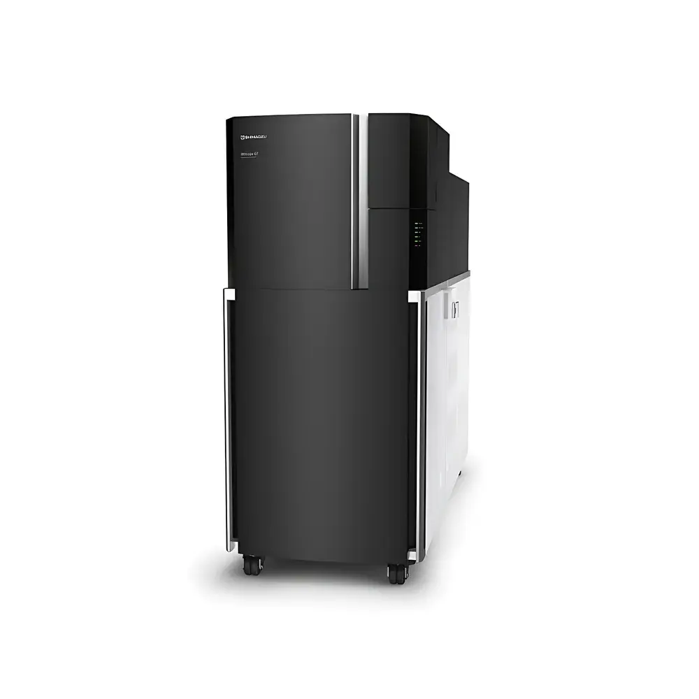
Shimadzu iMScope QT Imaging Mass Spectrometry Microscope
| Brand | Shimadzu |
|---|---|
| Origin | Japan |
| Manufacturer Type | Original Equipment Manufacturer (OEM) |
| Import Status | Imported |
| Model | iMScope QT |
| Instrument Type | MALDI-Q-TOF Hybrid Imaging System |
| Spatial Resolution | ≤5 µm |
| Sensitivity | Equivalent to LCMS-9030 |
| Mass Accuracy | <1 ppm over 24 h at ambient temperature |
| Acquisition Speed | ≥50 pixels/sec |
| Mass Precision | <1 ppm |
| Detection Platform | Integrated Optical Microscope–MALDI Source–Q-TOF Mass Spectrometer |
Overview
The Shimadzu iMScope QT is a fully integrated imaging mass spectrometry microscope engineered for spatially resolved molecular profiling of biological and material surfaces at cellular and subcellular resolution. It combines high-magnification optical microscopy with matrix-assisted laser desorption/ionization quadrupole time-of-flight (MALDI-Q-TOF) mass spectrometry in a single, co-registered platform. Unlike conventional off-line workflows requiring separate imaging and LC-MS analysis, the iMScope QT enables direct correlation of morphological features—observed in real time via its built-in high-resolution optical microscope—with molecular ion distributions mapped across the same sample surface. This co-localization capability is grounded in precise laser positioning, calibrated stage movement, and synchronized data acquisition between optical and mass spectral modalities. The system operates on the principle of raster-scanned MALDI ionization, where a focused UV laser (20 kHz repetition rate) ablates discrete microspots (down to 5 µm diameter) while the Q-TOF analyzer delivers high mass accuracy (<1 ppm), isotopic fidelity, and MS/MS structural elucidation—all without compromising spatial fidelity or analytical throughput.
Key Features
- Co-registered optical microscopy and MALDI-Q-TOF imaging in one instrument: Enables simultaneous visual inspection and molecular mapping with pixel-level alignment.
- Subcellular spatial resolution: Achieves ≤5 µm lateral resolution using optimized laser focus, delay-time gating, and high-precision XY stage control.
- High-speed imaging acquisition: Supports ≥50 pixels/sec acquisition rate when coupled with the LCMS-9030’s ultrafast TOF detection and high-duty-cycle ion optics.
- Modular architecture: The MALDI imaging unit and LCMS-9030 mass spectrometer are physically separable and reconfigurable—allowing rapid transition between imaging mode and high-sensitivity LC-Q-TOF liquid chromatography–mass spectrometry workflows.
- Mass accuracy stability: Maintains <1 ppm mass error over 24-hour continuous operation at ambient laboratory conditions (20–25 °C, 40–60% RH), meeting requirements for longitudinal biomarker tracking and cross-laboratory reproducibility.
- Full MS/MS capability: Leverages the LCMS-9030’s quadrupole precursor selection and high-transmission collision cell for targeted structural confirmation and specificity enhancement in imaging datasets.
Sample Compatibility & Compliance
The iMScope QT accepts standard MALDI-compatible substrates including conductive glass slides, ITO-coated slides, and metal targets. It supports frozen tissue sections (5–20 µm thickness), formalin-fixed paraffin-embedded (FFPE) sections (with antigen retrieval), cultured cells, plant tissues, and thin-layer chromatography plates. Sample preparation follows established MALDI protocols—including matrix deposition via robotic sprayer or sublimation—and is compatible with on-tissue derivatization for enhanced detection of low-abundance metabolites or lipids. From a regulatory standpoint, the system supports audit-trail-enabled data acquisition (aligned with FDA 21 CFR Part 11 requirements when used with compliant software configurations), and its mass accuracy and reproducibility meet ISO/IEC 17025 criteria for analytical method validation in clinical research and pharmaceutical development environments.
Software & Data Management
Data acquisition, processing, and visualization are managed through Shimadzu’s proprietary imScape software suite. imScape provides real-time optical–mass image overlay, peak alignment across multiple datasets, unsupervised clustering (e.g., k-means, PCA), and ion intensity normalization against internal standards or total ion current. Raw data files (.imzML compliant) are exportable for third-party analysis in SCiLS Lab, MSiReader, or Python-based platforms (e.g., Cardinal, PyImagingMS). All processing steps—including baseline correction, noise filtering, and peak picking—are logged with timestamped metadata, supporting GLP/GMP-compliant documentation. Batch processing pipelines support automated quantification across hundreds of regions-of-interest (ROIs), with optional integration into LIMS systems via standardized API interfaces.
Applications
- Neuroscience: Mapping neurotransmitter distribution, lipidomics gradients, and amyloid-beta peptide localization in brain tissue sections.
- Oncology research: Correlating tumor heterogeneity (morphology) with oncoprotein expression, drug metabolism, and resistance-associated metabolites in FFPE biopsies.
- Pharmaceutical development: Visualizing drug and metabolite penetration depth, distribution uniformity, and target engagement in preclinical tissue models.
- Plant science: Spatial profiling of secondary metabolites, phytohormones, and defense compounds across leaf vasculature or root–microbe interfaces.
- Single-cell and subcellular analysis: Resolving metabolic compartmentalization in isolated cells or organelle-enriched preparations using high-magnification objective lenses and sub-5 µm laser spots.
FAQ
Can the iMScope QT operate independently of the LCMS-9030?
Yes—the MALDI imaging module functions as a standalone unit with its own TOF detector; however, full Q-TOF performance (including MS/MS and sub-ppm mass accuracy) requires coupling to the LCMS-9030.
What is the minimum recommended sample size for optimal spatial resolution?
For ≤5 µm resolution, samples should be mounted flat on conductive substrates with minimal topographical variation; section thickness must not exceed 20 µm to prevent ion signal attenuation and delocalization.
Does the system support quantitative imaging?
Yes—via internal standard normalization, isotope-labeled spike-ins, or label-free relative quantification using TIC-normalized ion intensities; absolute quantitation requires calibration curves generated from adjacent tissue homogenates.
Is imScape software validated for regulated environments?
imScape v5.2+ includes electronic signature, audit trail, and user access control modules compliant with FDA 21 CFR Part 11 when deployed on validated Windows Server environments.
How is laser focus maintained during long-duration imaging runs?
The system employs an auto-focus routine triggered at user-defined intervals (e.g., every 1000 pixels) using reflected light feedback from the optical path, ensuring consistent ablation depth and signal reproducibility across multi-hour acquisitions.