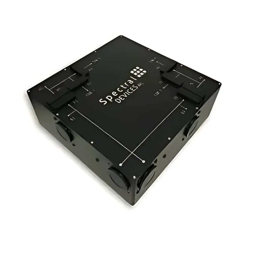
Spectral Devices Multi-Camera Imaging System (CMOS Global Shutter, 4-Channel)
| Origin | Canada |
|---|---|
| Manufacturer Type | Authorized Distributor |
| Origin Category | Imported |
| Model | CMOS Global Shutter |
| Price Range | USD 0–14,999 |
| Image Resolution | 2448 × 2048 |
| Pixel Size | 5.5 × 5.5 µm |
| Sensor Format | 1-inch |
| Maximum Readout Speed | 480 Mbps per channel |
| Dynamic Range | 60 dB |
Overview
The Spectral Devices Multi-Camera Imaging System is an engineered optical imaging platform designed for high-fidelity, simultaneous multi-spectral acquisition in advanced microscopy applications. Built upon a monolithic aluminum architecture and leveraging four synchronized CMOS global shutter sensors, the system splits a single incident optical path into four precisely calibrated, parallel imaging channels—each independently configurable with removable filter mounts and polarization optics. This optomechanical design eliminates temporal misalignment and spatial registration drift between channels, enabling true simultaneity in fluorescence imaging across distinct excitation/emission bands. Unlike time-multiplexed or scanning-based alternatives, the system captures four co-registered images from identical field-of-view (FOV) at full frame rate—critical for dynamic biological processes, FRET quantification, ratiometric calcium imaging, and multi-label immunofluorescence. The core measurement principle relies on spatially invariant beam splitting combined with hardware-level synchronization via optical trigger isolation, ensuring sub-microsecond inter-channel timing accuracy.
Key Features
- Four independent, co-aligned CMOS global shutter sensors (IMX249, CMV4000, or IMX250 variants) mounted on a thermally stable, CNC-machined 6061 aluminum chassis
- Native support for 1-inch optical filters and linear polarizers—enabling flexible spectral and polarization contrast configuration without external optics
- USB3 Vision and GenICam-compliant interface with real-time streaming up to 480 Mbps per channel; no external power supply required (bus-powered via USB 3.0)
- Hardware-triggered acquisition with galvanically isolated BNC I/O: one TTL-compatible trigger input and one synchronized output for external device coordination (e.g., laser pulsing, stage movement)
- Integrated cage-mount compatibility (24 × 4-40 threads) for seamless integration into 30 mm optical cage systems
- Anodized black surface finish with laser-etched labeling for traceability and ESD-safe operation in cleanroom-grade lab environments
Sample Compatibility & Compliance
The system is optimized for transmitted and epi-fluorescent microscopy configurations—including widefield, TIRF, and structured illumination setups—where spectral crosstalk, motion artifact, and photobleaching mitigation are paramount. It supports standard C-mount objective couplers and integrates directly with inverted and upright microscopes from Nikon, Olympus, Zeiss, and Leica. All firmware and host software comply with USB3 Vision v1.0 and GenICam SFNC 2.4 specifications. For regulated environments, the 2ndLook software framework supports audit trail logging, user access control, and metadata embedding compliant with ISO/IEC 17025 documentation requirements. While not FDA-cleared as a medical device, the system meets CE marking directives (2014/30/EU EMC and 2014/35/EU LVD) and RoHS 3 material restrictions.
Software & Data Management
The included 2ndLook for Windows application provides deterministic acquisition control, real-time preview, and synchronized multi-channel video recording in AVI, TIFF stack, and HDF5 formats—with optional lossless compression. It natively supports USB3 Vision, GigE Vision, and TWAIN-compliant external cameras, enabling hybrid acquisition workflows (e.g., combining the quad-camera head with a separate brightfield or IR camera). SDKs for C++, Python (via ctypes), and LabVIEW (NI Vision-compatible VIs) are provided for custom automation and integration into existing QC/QA pipelines. All captured frames include embedded EXIF-style metadata: exposure time, gain, sensor temperature (monitored via on-chip thermal diodes), timestamp (UTC-synchronized via host OS), and active filter ID. Data integrity is ensured through cyclic redundancy checks (CRC-32) on all transferred image buffers.
Applications
- Simultaneous four-color fluorescence imaging for multiplexed tissue staining (e.g., CD3/CD8/FOXP3/Ki67 in IHC/IF workflows)
- Ratiometric ion imaging (e.g., Ca2+, pH, Cl−) using dual-emission probes without mechanical filter wheels
- Live-cell dynamics tracking under low-light conditions—leveraging IMX249’s 1.3 e− read noise and 81 s maximum exposure for ultra-weak signal capture
- NIR-enhanced dual-band imaging (visible + 700–900 nm) using CMV4000-NIR variant for deep-tissue or scattering-sample applications
- Quantitative phase contrast + fluorescence correlation—by assigning one channel to differential interference contrast (DIC) and others to fluorophores
- Automated quality inspection in semiconductor metrology where multi-angle reflectance and defect classification require pixel-perfect channel alignment
FAQ
Does the system require external power?
No—the unit is fully bus-powered via its four USB 3.0 Type-A ports. Each camera channel draws ≤900 mA at 5 V DC, well within USB 3.0 specification limits.
Can I use third-party filters or custom dichroics?
Yes—the filter mounts accept standard 1-inch round optics (25.4 mm diameter, ≤6 mm thickness) with SM1-threaded retention rings. Custom dichroic cubes may be integrated using optional adapter plates.
Is Linux driver support available?
Yes—GenICam-compliant drivers for Ubuntu 20.04+ and CentOS 7.x are included, along with sample code for Aravis and Vimba SDK integration.
What is the maximum sustained frame rate across all four channels?
At full resolution (2448 × 2048), the system achieves 36 fps per channel (144 total streams) with 12-bit depth over aggregated USB 3.0 bandwidth. ROI-based acquisition enables >1,000 fps per channel on CMV4000 variants.
How is inter-channel geometric calibration maintained?
Each optical path undergoes factory-applied affine transformation mapping using NIST-traceable grid targets. Calibration matrices are stored in non-volatile memory and applied in real time during acquisition—no user recalibration required under normal thermal operating range (15–30 °C).


