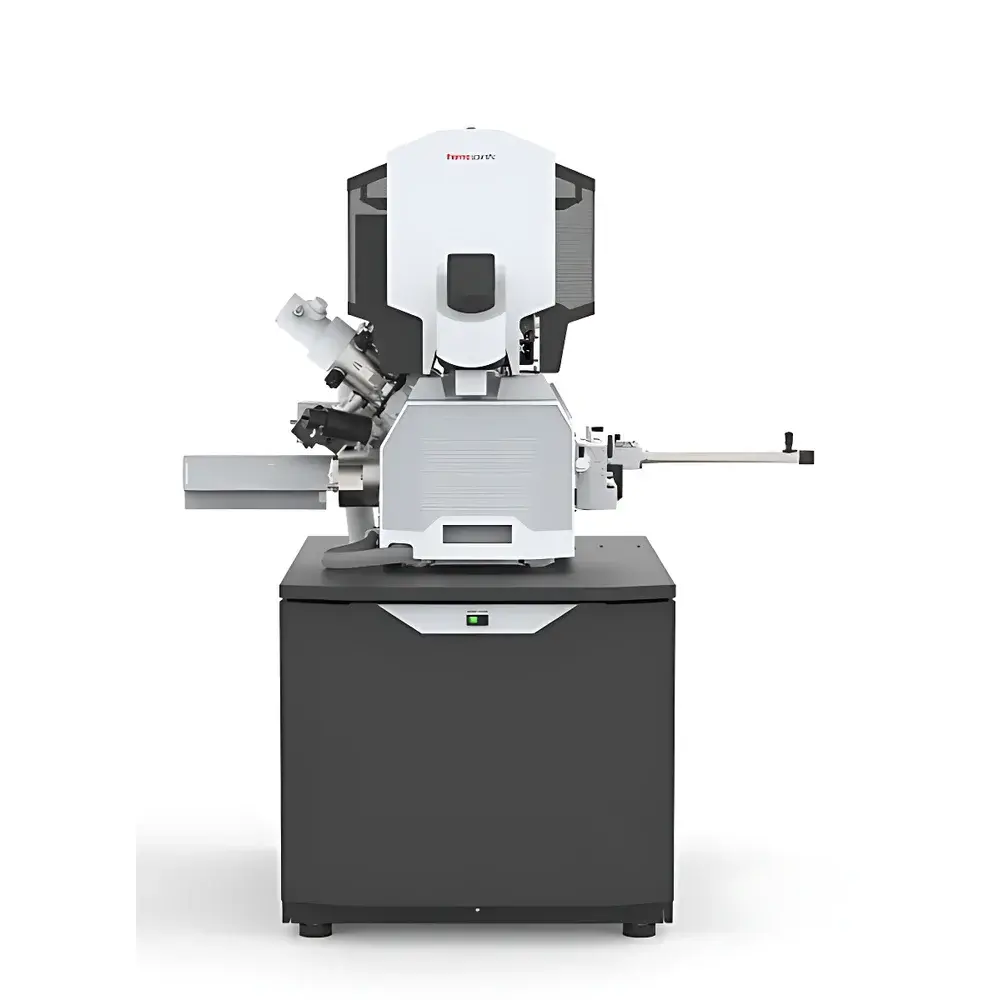
Thermo Fisher Scientific Aquilos 2 Cryo-Focused Ion Beam (Cryo-FIB) Microscope
| Brand | Thermo Fisher Scientific |
|---|---|
| Origin | Imported |
| Manufacturer Type | Original Equipment Manufacturer (OEM) |
| Model | Aquilos 2 Cryo-FIB |
| Price Range | USD 1.3–2.0 million (FOB) |
Overview
The Thermo Fisher Scientific Aquilos 2 Cryo-Focused Ion Beam (Cryo-FIB) Microscope is a purpose-built, dual-beam cryogenic platform engineered for high-fidelity lamella preparation in cryo-electron tomography (cryo-ET). Unlike conventional room-temperature FIB systems, the Aquilos 2 operates exclusively under cryogenic conditions—maintaining samples at ≤−185 °C throughout imaging, milling, and transfer—to preserve native hydration states and prevent devitrification or ice crystallization. Its core functionality relies on gallium ion beam sputtering under ultra-high vacuum (UHV) and cryo-stable conditions, enabling precise, artifact-free thinning of vitrified biological specimens directly on TEM grids. The system integrates seamlessly with high-end cryo-transmission electron microscopes (cryo-TEMs), serving as the critical upstream sample preparation module in structural cell biology workflows where molecular-scale fidelity is non-negotiable.
Key Features
- Cryo-optimized dual-beam architecture combining a field-emission scanning electron microscope (SEM) and a liquid-metal Ga⁺ focused ion beam (FIB) column, both aligned and calibrated for sub-10 nm positional accuracy at cryogenic temperatures.
- Full-rotation cryo-stage (±90° tilt, 360° rotation) with active cooling via closed-cycle helium refrigeration, ensuring thermal stability during extended milling sequences.
- Dedicated cryo-autoloader compatible with standard Gatan 626/656 cryo-transfer holders and Thermo Fisher’s AutoGrid™ carriers—enabling unattended grid loading, alignment, and stage positioning.
- Integrated anti-contamination cold trap and cryo-shielded beam pathways to minimize ice deposition on the sample surface during milling; typical ice growth rates <0.5 nm/min under operational vacuum (<1×10⁻⁷ mbar).
- Automated lamella workflow powered by Thermo Fisher’s Maps™ software suite, including guided lamella targeting, real-time thickness monitoring via ion-induced secondary electron contrast, and endpoint detection based on electron transparency thresholds.
- Robust mechanical design compliant with ISO 14644-1 Class 5 cleanroom requirements for cryo-chamber housing, reducing particulate-induced artifacts during long-duration runs.
Sample Compatibility & Compliance
The Aquilos 2 supports a broad range of cryo-preserved biological specimens—including cultured mammalian cells, primary neurons, bacterial biofilms, and isolated organelles—mounted on standard Au or Ni cryo-EM grids (e.g., Quantifoil R2/2, UltrAuFoil). All hardware and software modules are designed to meet GLP-compliant documentation standards, supporting audit-ready operation logs, user-access controls, and timestamped parameter tracking per session. The system adheres to ISO/IEC 17025 principles for method validation in analytical laboratories and is compatible with FDA 21 CFR Part 11–enabled electronic record systems when integrated with Thermo Fisher’s TFS Connect™ enterprise software infrastructure.
Software & Data Management
Maps™ software provides a unified interface for correlative light–electron microscopy (CLEM) integration: optical fluorescence data (e.g., from widefield or confocal microscopes) can be imported as overlays onto SEM/FIB navigation maps, enabling precise targeting of fluorescently labeled subcellular structures. All acquisition parameters—including beam currents, dwell times, milling angles, and stage coordinates—are stored in vendor-neutral HDF5 format with embedded metadata compliant with EMDB and PDB standards. Automated report generation includes lamella geometry metrics (thickness, width, taper angle), milling duration, and vacuum history—exportable for LIMS integration or regulatory submission packages.
Applications
- Preparation of sub-100 nm-thick cryo-lamellae from intact eukaryotic cells for high-resolution cryo-ET reconstruction of macromolecular complexes in situ.
- Routine lamella fabrication from thick (>10 µm) frozen-hydrated tissue sections, including brain slices and organoids, without compression artifacts inherent to mechanical sectioning.
- Site-specific extraction of intracellular targets (e.g., synaptic vesicles, nuclear pore complexes) using cryo-FIB lift-out combined with in-situ TEM transfer.
- Correlative structural analysis across modalities: correlating super-resolution fluorescence localization with 3D cryo-ET volumes to map protein distribution within cellular ultrastructure.
- Method development for cryo-FIB milling optimization, including dose-controlled low-kV ion beam trimming and multi-angle milling strategies to reduce curtaining effects.
FAQ
What is the minimum achievable lamella thickness using the Aquilos 2?
Typical final lamella thickness ranges from 50–120 nm, depending on specimen density and ion beam energy; routine reproducibility ±5 nm is attainable under optimized conditions.
Is the Aquilos 2 compatible with third-party cryo-TEMs?
Yes—the system uses standard cryo-transfer protocols (Gatan 626/656, FEI/TFS Titan-compatible carriers) and supports manual or automated handoff to most commercial cryo-TEM platforms.
Does the system support automated overnight lamella production?
Yes—fully programmed workflows, including grid mapping, target identification, rough milling, fine milling, and polishing, can execute unattended for up to 16 hours with continuous vacuum and temperature monitoring.
How does the Aquilos 2 mitigate ion beam damage during milling?
Through synchronized low-dose imaging, variable kV ion beam ramping (5–30 keV), and cryo-stabilized stage dynamics that suppress radiolytic heating and mass loss—validated via EELS and EDX line scans showing minimal elemental redistribution.
Can Maps software import data from non-Thermo Fisher optical microscopes?
Yes—TIFF, OME-TIFF, and ND2 formats are natively supported; spatial registration uses fiducial-based affine transformation with sub-pixel accuracy.


