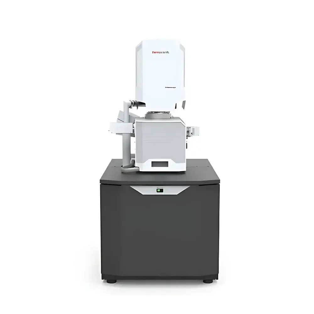
Thermo Scientific Volumescope 2 Scanning Electron Microscope
| Brand | Thermo Fisher Scientific |
|---|---|
| Origin | Imported |
| Manufacturer Type | Original Equipment Manufacturer (OEM) |
| Model | Volumescope 2 |
| Instrument Type | Floor-standing SEM |
| Electron Source | Cold Field Emission Gun (CFEG) |
| Accelerating Voltage | 1.0–30 kV |
| Vacuum Modes | High Vacuum (HiVac) and Low Vacuum (LoVac) with VSDBS detector |
| Z-axis Optical Sectioning Resolution | Up to 10 nm |
| Sample Handling | Automated ultramicrotome integration for seamless transition between conventional SEM and SBFI/AT workflows |
Overview
The Thermo Scientific Volumescope 2 is a purpose-built, floor-standing scanning electron microscope engineered specifically for high-fidelity, large-volume 3D ultrastructural analysis in life sciences research. Unlike conventional SEMs optimized for surface topography, the Volumescope 2 integrates a synchronized in-chamber ultramicrotome and cold field emission electron source to enable automated serial block-face imaging (SBFI)—a technique that combines precise physical sectioning with sequential high-resolution backscattered electron (BSE) imaging. This architecture ensures isotropic spatial resolution across x-, y-, and z-dimensions, with verified optical sectioning capability down to 10 nm in the z-axis—critical for reconstructing faithful volumetric models of neural circuits, organelle networks, or tissue microenvironments. Its design adheres to rigorous engineering standards for thermal and mechanical stability, minimizing drift during extended acquisitions (hours to days), and supports both high-vacuum (HiVac) and low-vacuum (LoVac) imaging modes to accommodate diverse biological specimen requirements without metal coating.
Key Features
- Cold field emission gun (CFEG) delivering high-brightness, stable electron beam with sub-5 nm probe size at 30 kV—enabling high signal-to-noise ratio imaging at nanoscale resolution.
- Integrated in-chamber ultramicrotome with sub-10 nm sectioning repeatability, synchronized with BSE detection for artifact-minimized serial imaging.
- Dual vacuum operation: HiVac mode for conductive or metal-coated samples; LoVac mode with Variable Pressure Secondary Electron Detector (VSDBS) for uncoated, beam-sensitive, or moderately charging biological specimens—maintaining comparable contrast and resolution to HiVac.
- Amira Live Tracker software module enabling real-time ROI navigation, multi-region acquisition planning, and on-the-fly adjustment of imaging parameters during unattended runs.
- Automated sample handling interface supporting rapid exchange between standard SEM imaging and SBFI/Array Tomography (AT) workflows—facilitated by optional Thermo Scientific Maps software for correlative light-electron microscopy (CLEM) registration and multiplexed AT alignment.
- Fragment detection and Swipe functionality: algorithm-driven identification of section debris followed by automated stage repositioning and slice discard—preserving data integrity and protecting irreplaceable samples from contamination or misalignment.
Sample Compatibility & Compliance
The Volumescope 2 accommodates a broad spectrum of life science specimens—including resin-embedded brain tissue blocks (e.g., epoxy or acrylic), cryo-sectioned samples (with optional cryo-stage), and semi-thick plastic sections (50–200 nm). Its LoVac/VSDBS configuration meets common requirements for charge mitigation in hydrated or lightly stained samples per ASTM E1558 and ISO 16700 guidelines for SEM-based biological imaging. System firmware and acquisition logs comply with GLP-aligned audit trail requirements, supporting traceability for preclinical and translational research environments. All hardware and software components are CE-marked and conform to IEC 61000-6-3 (EMC) and IEC 61010-1 (safety) standards.
Software & Data Management
Acquisition and reconstruction are managed through Thermo Scientific Avizo and Amira software platforms, supporting DICOM-compliant export, HDF5-based large-volume data storage, and GPU-accelerated 3D rendering. The system generates FAIR-compliant metadata (Findable, Accessible, Interoperable, Reusable) including acquisition parameters, stage coordinates, and vacuum conditions embedded directly in image headers. For regulated environments, optional 21 CFR Part 11 compliance packages provide electronic signature support, role-based access control, and immutable audit logs for all user actions—from parameter modification to dataset export.
Applications
- Connectomics: Reconstruction of synaptic connectivity maps in mouse, primate, and human brain tissue at synaptic-level resolution.
- Cellular ultrastructure: 3D mapping of mitochondrial dynamics, ER morphology, and vesicle trafficking within intact cells.
- Tissue pathology: Volumetric assessment of tumor microarchitecture, immune cell infiltration patterns, and vascular network remodeling in FFPE or resin-embedded biopsies.
- Developmental biology: Time-series SBFI of embryonic organogenesis using serially sectioned whole-mount samples.
- Correlative multimodal imaging: Integration with fluorescence light microscopy (via Maps CLEM) to overlay functional protein localization onto ultrastructural context.
FAQ
What vacuum modes does the Volumescope 2 support, and when should each be used?
The system operates in High Vacuum (HiVac) for optimal resolution on conductive or metallized samples, and Low Vacuum (LoVac) with the VSDBS detector for uncoated, beam-sensitive, or moderately charging biological specimens—without compromising contrast or z-resolution.
Is the ultramicrotome fully integrated into the SEM chamber?
Yes—the in-chamber ultramicrotome is mechanically and electronically synchronized with the electron column and stage, enabling sub-pixel registration between sectioning and imaging steps.
Can the Volumescope 2 perform Array Tomography (AT)?
AT is supported as an optional workflow using Thermo Scientific Maps software, which enables automated multi-channel fluorescence registration and alignment of serial sections imaged by TEM or SEM.
How does the system protect valuable samples during long acquisitions?
Through fragment detection algorithms, automated swipe routines, real-time drift correction, and dual-mode vacuum control—minimizing beam damage, charging artifacts, and mechanical contamination over multi-day runs.
What level of z-axis resolution is achievable in SBFI mode?
Optical sectioning resolution is validated at ≤10 nm in the z-direction under standard operating conditions, ensuring true isotropic voxel sampling for quantitative 3D morphometry.


