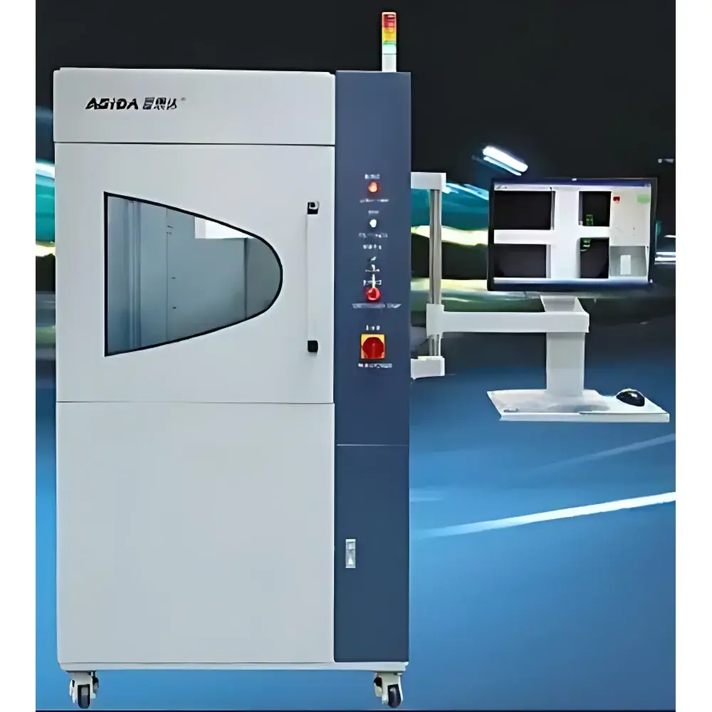
XG5000 Battery Defect Detection System – Microfocus X-ray Imaging Instrument
| Origin | Guangdong, China |
|---|---|
| Manufacturer Type | Authorized Distributor |
| Origin Category | Domestic (PRC) |
| Model | XG5000 |
| Pricing | Upon Request |
| X-ray Tube | Sealed microfocus, integrated high-voltage supply |
| Max. Tube Voltage | 90 kV |
| Max. Tube Current | 200 µA (software-limited to 89 µA) |
| Cooling | Forced air |
| Geometric Magnification | 12×–48× |
| Detectable Feature Size | ≥5 µm |
| X-Y-Z Travel Range | 400 mm × 450 mm × 150 mm |
| Image Intensifier FOV | 2″ / 4″ selectable |
| Resolution | 75–110 lp/cm |
| Radiation Leakage | ≤1 µSv/hr at 5 cm from enclosure |
| Operating Temp. | 22 ± 3 °C |
| Power Supply | AC 250 V, 10 A |
| System Weight | ~760 kg |
| Compliance | Meets IEC 61331-1, IEC 62495, and GBZ 138–2019 radiation safety requirements |
Overview
The XG5000 Battery Defect Detection System is a benchtop microfocus X-ray imaging platform engineered for non-destructive internal inspection of electrochemical energy storage devices. It operates on the principle of transmission radiography: a sealed, integrated 90 kV microfocus X-ray tube generates a highly collimated beam that penetrates battery cells—cylindrical, prismatic, or pouch-type—while an image intensifier captures differential attenuation across structural features. This enables high-contrast visualization of electrode delamination, dendrite formation, separator wrinkles, weld voids, tab misalignment, and foreign particle inclusions down to 5 µm in equivalent density contrast. Designed specifically for QC laboratories and R&D centers in lithium-ion battery manufacturing, the system delivers quantitative geometric magnification up to 48× with sub-micron spatial registration repeatability, supporting root-cause analysis during failure mode investigation (FMEA), process validation, and incoming material screening.
Key Features
- Sealed microfocus X-ray tube with integrated high-voltage power supply (90 kV, 200 µA max), eliminating external HV cabling and enhancing long-term stability.
- Precision motion control via ball-screw-driven XYZ stages (400 mm × 450 mm × 150 mm travel), synchronized stepper motors, and low-noise mechanical architecture for repeatable positioning accuracy ≤±2 µm.
- Automated tube protection: system enters standby after 20 minutes of inactivity; immediate beam termination upon interlock trigger (e.g., door opening), compliant with Class I radiation safety requirements per IEC 61331-1.
- Real-time navigational imaging: click-to-center functionality with dynamic crosshair overlay; stored coordinate recall with associated exposure parameters (kV, µA, integration time) for multi-point comparative analysis.
- On-screen measurement toolkit: calibrated linear distance, point-to-line, circle diameter, concentricity, and radial offset calculations—all traceable to NIST-traceable calibration standards.
- Intuitive GUI with color-coded X-ray status indicator (green = active, red = off), mouse-wheel zoom/pan, and persistent scale bar overlay for rapid dimensional assessment without post-processing.
Sample Compatibility & Compliance
The XG5000 accommodates standard commercial battery formats up to 300 mm in height and 200 mm in width—including 18650, 21700, 26650 cylindrical cells; 50–120 mm wide prismatic modules; and flexible pouch cells mounted on rigid carriers. Sample handling is compatible with ISO 17025-accredited workflows: all exposure parameters are logged with timestamps and user IDs, supporting audit-ready documentation for GLP/GMP environments. Radiation shielding meets IEC 62495 (Safety of Industrial X-ray Equipment) and GBZ 138–2019 (Chinese National Standard for X-ray Equipment Protection), with verified leakage ≤1 µSv/hr at 5 cm from any surface. Optional tilt-stage and auto-navigation modules extend capability for angled tomographic projection sequences under controlled protocol.
Software & Data Management
The proprietary acquisition and analysis software runs natively on Windows OS (Windows 10 LTSB recommended; backward-compatible with Windows 7/XP). All image metadata—including tube voltage, current, focal spot size, geometric magnification factor, stage coordinates, and detector gain settings—is embedded in DICOM-compliant headers. Measurement results export directly to CSV or Excel for statistical process control (SPC) integration. Audit trail functionality records operator logins, parameter changes, image annotations, and report generation events—fully compliant with FDA 21 CFR Part 11 requirements when deployed with validated electronic signature modules. Raw image archives support lossless TIFF and compressed JPEG2000 formats; batch processing scripts enable automated defect thresholding and pass/fail flagging based on user-defined grayscale deviation criteria.
Applications
- Root-cause analysis of thermal runaway precursors: detection of metallic burrs, electrode folding, and localized separator thinning.
- Weld integrity verification in busbar and tab connections—identifying voids, cracks, and insufficient fusion zones.
- Separator integrity assessment: wrinkling, pore collapse, and shrinkage behavior under thermal stress simulation.
- Post-cycling degradation tracking: quantifying anode swelling, cathode cracking, and electrolyte dry-out patterns across charge/discharge cycles.
- Supplier qualification: comparative imaging of raw electrode foils and pre-assembled jellies to enforce incoming material specifications aligned with UL 1642 and IEC 62133-2 test readiness protocols.
FAQ
What is the minimum detectable defect size under optimal conditions?
Under nominal 90 kV, 89 µA exposure with 48× geometric magnification and 2″ FOV, the system resolves features ≥5 µm in equivalent density contrast (e.g., voids in copper foil or Ni-rich cathode layers). Actual resolution depends on sample thickness, material composition, and signal-to-noise ratio.
Is the system suitable for in-line production use?
The XG5000 is optimized for laboratory-based QA/QC and R&D—not high-throughput inline deployment. Its programmable step-and-repeat function supports semi-automated batch inspection of uniform geometries, but cycle time remains >30 s per scan due to mechanical stabilization and image integration requirements.
Does the software support automated defect classification?
Baseline software provides manual annotation and threshold-based region-of-interest (ROI) analysis. AI-assisted defect classification (e.g., CNN-based anomaly detection) is available as an optional module requiring separate validation per ISO/IEC 17025 clause 5.4.2 for algorithmic measurement systems.
How is radiation safety ensured during routine operation?
The fully interlocked lead-shielded enclosure includes redundant door switches, real-time dose monitoring, and automatic beam cutoff within <100 ms of interlock breach. Annual third-party radiation survey certification is recommended per national regulatory authority requirements.
Can the system be upgraded for computed tomography (CT)?
Yes—optional rotary stage integration (with ±0.01° angular resolution) and CT reconstruction software license enable full 3D volumetric rendering. CT mode requires additional shielding evaluation and compliance re-certification per IEC 61223-3-5.
Related Products


")