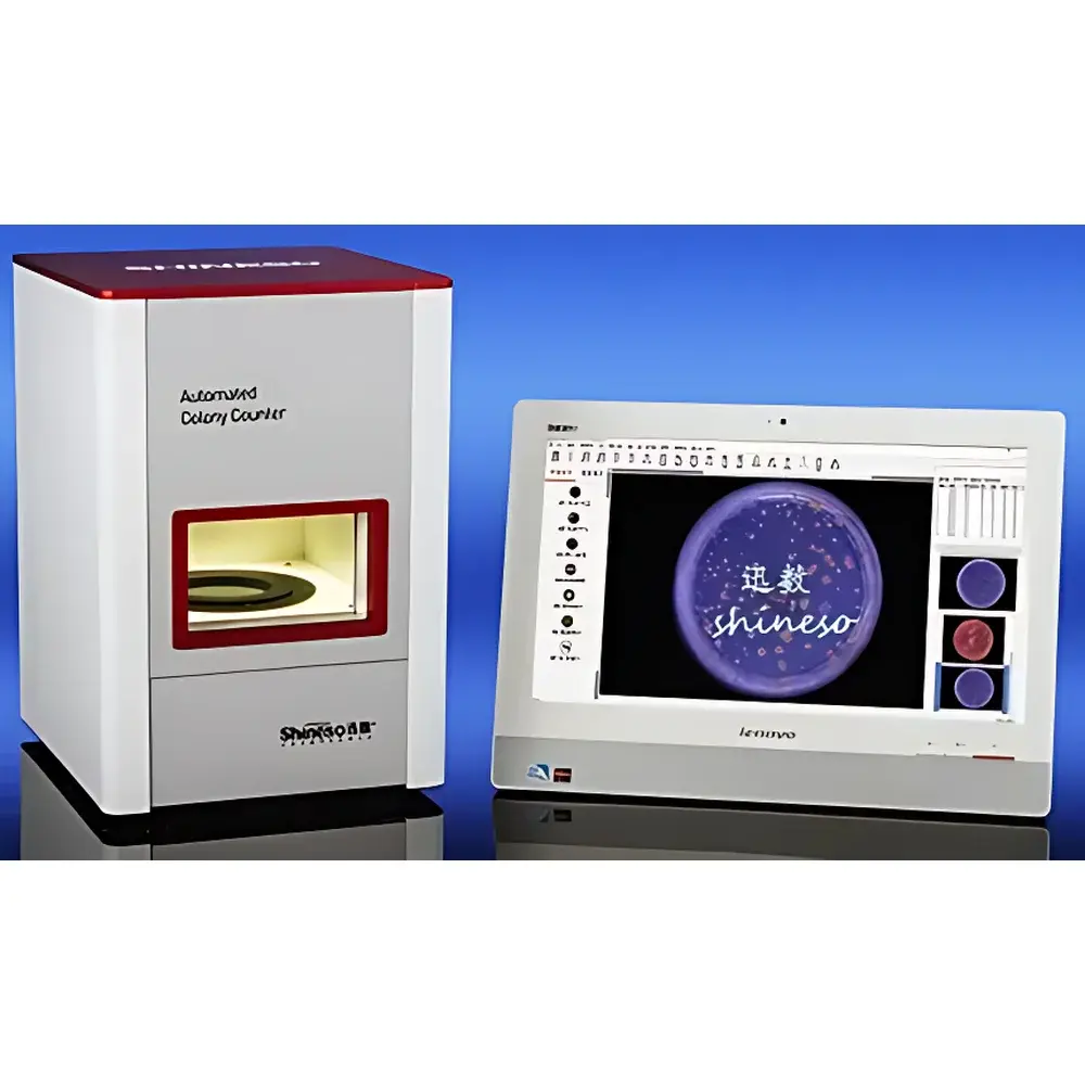
Xunshu iCount 33 Fully Automated Colony Counter
| Brand | Xunshu |
|---|---|
| Origin | Zhejiang, China |
| Manufacturer Type | Original Equipment Manufacturer (OEM) |
| Instrument Type | Fully Automated Colony Counter |
| Sample Types | Liquid media, solid media (pour plate, spread plate, membrane filtration, spiral plating, commercial test plates) |
| Petri Dish Capacity | 35–150 mm diameter |
| Counting Speed | 20,000 colonies/sec |
| Resolution | 0.01 mm |
| Imaging System | Fixed-focus 8 mm lens, 5.0 MP, 2/3″ optical format |
| CMOS sensor size | 1/2.4″, physical resolution: 8.5 megapixels (3328 × 2548), pixel size: 1.67 × 1.67 µm |
| Configuration | Main unit + colony analysis software suite |
Overview
The Xunshu iCount 33 Fully Automated Colony Counter is a fifth-generation microbiological imaging and enumeration system engineered for precision, reproducibility, and operational efficiency in regulated and research laboratory environments. It employs digital image acquisition coupled with multi-modal illumination physics—specifically diffuse reflective top lighting, tunable transmissive backlighting, and dual-wavelength suspended dark-field illumination—to resolve morphologically diverse microbial colonies across heterogeneous agar surfaces. Unlike conventional colony counters relying on single-spectrum or fixed-angle illumination, the iCount 33 implements a tri-illuminant optical architecture that enables high-fidelity contrast generation under variable colony pigmentation, transparency, and substrate opacity conditions. Its core measurement principle is based on high-resolution monochrome and color segmentation of digitized petri dish images, followed by morphometric classification using adaptive thresholding, level-set segmentation, and feature-based clustering algorithms—all compliant with ISO 4833-1:2013 (microbiology of food and animal feeding stuffs — horizontal method for the enumeration of microorganisms — Part 1: Colony count at 30 °C) and ASTM E2924-21 (standard guide for validation of digital image analysis systems for microbiological enumeration). Designed for walk-up usability without sacrificing analytical rigor, the system supports both routine QC workflows and advanced research applications requiring traceable, auditable, and repeatable colony quantification.
Key Features
- Fully enclosed aluminum-alloy chassis (32 × 34 × 46 cm) ensuring optical isolation, mechanical stability, and UV containment (254 nm internal lamp for chamber decontamination)
- Tri-illuminant optical engine: (1) Embedded 96-LED diffuse reflectance system with nanoscale light-scattering material (3100–5800 K CCT, 50–7000 lux adjustable); (2) Patented “Ling-Tou” white LED transmissive backlight with directional control for enhanced depth perception of translucent colonies; (3) Dual-wavelength (white + blue) suspended dark-field illumination generating high-contrast halo effects against deep-blue backgrounds
- Industrial-grade imaging subsystem: C-mount 8 mm fixed focal length lens (distortion <1%, f/1.4–f/16), 8.5 MP CMOS sensor (3328 × 2548), 1.67 µm pixel pitch, USB 3.0 interface for sub-second frame capture
- Intuitive parameter-free statistical interface: Scroll-wheel–driven real-time adjustment across four optimized counting modes (homogeneous background, uneven illumination, microcolonies, colored substrates) plus three one-click protocols (monochrome, mold/yeast, inverse contrast)
- Advanced segmentation algorithms including level-set multi-model optimization, bias-estimation clustering, and hue-saturation-value (HSV)-guided selective colony recognition for mixed-culture plates
- Comprehensive contamination management tools: manual region exclusion, grid-line removal, marker ink suppression, and user-guided splitting of clustered or filamentous colonies
Sample Compatibility & Compliance
The iCount 33 accommodates standard and non-standard microbiological sample formats—including pour plates, spread plates, membrane filters (black-grid and plain), spiral-plated dishes, and commercially available test kits (3M Petrifilm™ series for total aerobic count, Staphylococcus aureus, coliforms, and E. coli/coliform rapid detection). Its illumination adaptability ensures reliable detection of low-contrast colonies on chromogenic media (e.g., VRBA, Rose Bengal Agar) and metabolically active colonies on TTC-impregnated films. All data handling conforms to GLP and GMP principles: electronic audit trails record operator ID, timestamp, image acquisition parameters, algorithm selection, manual corrections, and dilution factor inputs. The software architecture supports 21 CFR Part 11–compliant user authentication, role-based access control (“Manager”, “Operator”, “Reviewer”), and immutable PDF/Excel export with embedded metadata and image thumbnails.
Software & Data Management
The proprietary colony analysis software provides a modular, hierarchical workflow—from raw image import to final report generation. Core modules include automatic calibration (reference bead or stage micrometer), dynamic ROI definition (circular, rectangular, polygonal, or “cut-out” multi-zone), and granular morphometric analysis (equivalent diameter, area, perimeter, aspect ratio, circularity, Feret’s diameter). Statistical outputs are automatically normalized per ISO 4833-1 requirements: colony counts are reported as CFU/mL or CFU/g after inputting dish diameter and sample dilution factor. All session data—including annotated images, intermediate segmentation masks, and parameter logs—are stored in a relational SQLite database with optional network backup. Export options include ANSI-compliant CSV, ISO 22000–aligned PDF reports with header/footer branding, and Excel-compatible .xlsx files containing full colony-by-colony attribute tables.
Applications
The iCount 33 serves laboratories engaged in food safety testing (AOAC-certified methods for pathogen screening), pharmaceutical environmental monitoring (ISO 14644-1 classified cleanrooms), clinical microbiology (urine, wound, and sputum culture analysis), water quality assessment (membrane filtration standards per EPA Method 1604), and academic research involving fungal sporulation kinetics or antibiotic susceptibility gradient assays. Its ability to distinguish subtle phenotypic variations makes it suitable for strain differentiation studies where colony texture, edge morphology, and pigment diffusion patterns serve as taxonomic indicators. In regulatory settings, its deterministic image processing pipeline supports method validation per ICH Q5C and USP .
FAQ
Does the iCount 33 support FDA 21 CFR Part 11 compliance?
Yes—the software implements electronic signatures, audit trail logging, role-based permissions, and immutable data archiving to meet Part 11 requirements for electronic records and signatures.
Can the system process 3M Petrifilm™ test plates without manual intervention?
Yes—dedicated one-click modules recognize and enumerate colonies on 3M Total Aerobic Count, S. aureus, Coliform, and E. coli/Coliform Rapid tests, including auto-detection of indicator dye reactions and inhibition zones.
What is the minimum resolvable colony size?
The system achieves 0.01 mm spatial resolution at 150 mm dish diameter, enabling reliable detection of colonies ≥100 µm in diameter under optimal contrast conditions.
Is external calibration required before each use?
No—the built-in calibration routine uses a standardized reference target; however, periodic verification with NIST-traceable stage micrometers is recommended per ISO/IEC 17025 quality assurance protocols.
How does the system handle overlapping or chain-forming colonies?
It applies watershed-based splitting with user-adjustable separation intensity, plus optional manual intervention via polygonal splitting tools—fully documented in the audit trail.
Can results be integrated into LIMS platforms?
Yes—CSV and XML exports are structured for direct ingestion into common LIMS architectures; API documentation is available upon request for custom integration.


