Xunsu A100 Photostaining Microplate Imaging Analyzer & Colony Counter
| Brand | Xunsu |
|---|---|
| Origin | Zhejiang, China |
| Manufacturer Type | Original Equipment Manufacturer (OEM) |
| Product Origin | Domestic (China) |
| Model | A100 |
| Pricing | Upon Request |
Overview
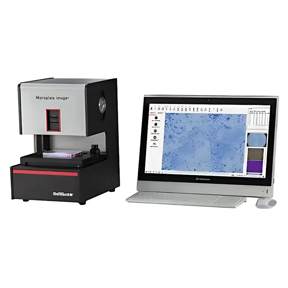
The Xunsu A100 Photostaining Microplate Imaging Analyzer & Colony Counter is a high-content, macro-scale digital imaging platform engineered for quantitative analysis of adherent and suspended cellular structures across standard multiwell plates (6–96-well), Petri dishes (35 mm), and specialized substrates including Transwell inserts, ELISPOT membranes, and Ames test microplates. It employs a hybrid optical architecture combining wide-field trans-illumination,悬浮-style darkfield illumination, and patented Rheinberg photostaining—where spectrally tunable excitation light couples with color-filtered backlighting to generate high-contrast, label-free optical contrast in unstained or lightly stained colonies, plaques, or biofilms. This enables precise morphometric quantification without reliance on chemical fixation or enzymatic development—critical for preserving sample integrity in longitudinal assays and GLP-compliant workflows. Designed for reproducible macro-imaging under controlled illumination conditions, the A100 delivers full-well, single-shot acquisition with sub-10 µm spatial resolution at 96-well scale, supporting both qualitative documentation for publication and quantitative image-based cytometry per ISO/IEC 17025 and ASTM E2925 guidelines.
Key Features
- Triple-mode illumination system: adjustable darkfield (100–5500 lux, CRI ≥74), color-tunable brightfield (11-channel LED backlight, uniformity >93%), and Rheinberg photostaining (multi-spectral excitation + wide-field chromatic backlighting)
- Motorized macro-zoom lens with calibrated field-of-view presets for 6-, 12-, 24-, 48-, and 96-well plates—enabling true full-well, non-mosaic imaging in one exposure
- 1.1-inch global-shutter CMOS sensor (Sony IMX series), 10+ megapixels, dual-layer noise reduction, and real-time depth-fusion stacking for extended-focus imaging of vertically distributed colonies
- Integrated mechanical stage with precision positioning for repeatable plate registration; compatible with standard SBS-format microplates, 35 mm dishes, slides, hemocytometers, and Transwell inserts
- Embedded optical alignment system: rotary-sliding light-field locator for rapid source repositioning, built-in iris diaphragm to suppress stray light, and harmonic-gear coarse/fine focus mechanism for drift-free stability
Sample Compatibility & Compliance
The A100 supports diverse life science applications requiring regulatory traceability and analytical rigor. It is routinely deployed in OECD 471-compliant Mini-Ames assays, USP -aligned microbial susceptibility testing (MIC determination via broth microdilution), and FDA-recognized clonogenic survival assays per ICH S9. Its software architecture includes audit-trail logging, user-role-based access control (administrator/operator tiers), and export compliance with 21 CFR Part 11 requirements—including electronic signatures, time-stamped metadata, and immutable raw image archiving. All image processing operations are fully documented and reversible, supporting GxP validation protocols for QC labs and preclinical research units operating under ISO 9001 or ISO/IEC 17025 frameworks.
Software & Data Management
The bundled Xunsu CellVision Suite comprises two modular applications: Microplate Colony Analyzer (MCA) and Multi-Modal Cell Imaging Analyzer (MCIA). MCA provides automated colony detection using seven complementary segmentation algorithms—adaptive thresholding, watershed separation, shape-based filtering, intensity gradient mapping, and deep-learning-assisted contour refinement—to distinguish overlapping clones, microcolonies, or plaque variants under variable contrast conditions. MCIA extends functionality to scratch-wound healing kinetics, confluence estimation via level-set segmentation, fluorescence channel unmixing (Hoechst/PI/CFSE), and ELISPOT spot quantification with halo-aware centroid detection. Data output includes Excel-compatible spreadsheets with per-colony metrics (equivalent diameter, area, perimeter, circularity, optical density, volume proxy), batch-exportable TIFF/PDF reports, and DICOM-compliant metadata embedding for LIMS integration.
Applications
- Clonogenic assays: soft-agar colony formation, monolayer colony survival, and stem cell sphere quantification
- Virology: plaque assay (crystal violet/neutral red-stained), TCID50 endpoint titration, and viral cytopathic effect scoring
- Immunology: ELISPOT enumeration with automatic spot sizing, halo intensity profiling, and background subtraction
- Toxicology & mutagenicity: Mini-Ames and Micro-Ames assay imaging, mouse lymphoma L5178Y TK+/− mutation scoring, and HGPRT gene mutation analysis
- Cell migration/invasion: full-membrane Transwell imaging with membrane-edge registration and bottom-surface cell counting
- Stem cell differentiation: ALP and Alizarin Red S staining quantification, osteogenic nodule classification, and spindle-cell morphology indexing
- In vivo tumor modeling: subcutaneous xenograft measurement (length/width/volume calculation from dorsal-view images)
- Microbial analysis: MIC endpoint determination via turbidity grading, biofilm photostaining, and microcolony morphology characterization
FAQ
Does the A100 require fluorescent dyes for colony visualization?
No—Rheinberg photostaining enables label-free optical contrast enhancement through spectral interference between excitation and backlight channels, eliminating the need for chemical staining in many applications.
Can the system validate against pharmacopeial standards such as USP ?
Yes—the illumination uniformity (>93%), spatial resolution (≤8 µm at 96-well), and software audit trail capabilities align with USP verification criteria for image-based microbiological methods.
Is the software compliant with 21 CFR Part 11 for regulated environments?
Yes—user authentication, electronic signature capture, immutable raw data archiving, and change-controlled versioning meet core Part 11 requirements for electronic records and signatures.
What is the maximum field-of-view resolution for a 6-well plate?
At 6-well scale, the system achieves ≤20 µm/pixel resolution with full-well single-shot acquisition, preserving morphological fidelity without stitching artifacts.
Does the A100 support time-lapse imaging?
While optimized for endpoint macro-imaging, scheduled batch acquisition with timestamped metadata enables semi-automated longitudinal monitoring across multiple plates over defined intervals.


