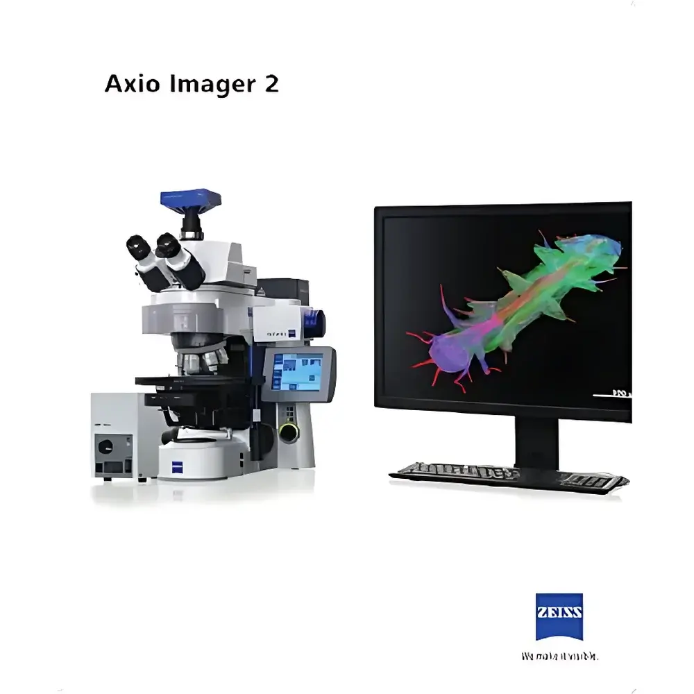
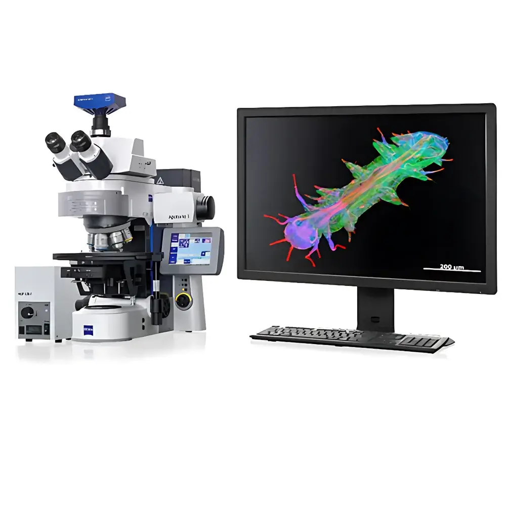
ZEISS Axio Imager 2 Research-Grade Upright Microscope
| Brand | ZEISS |
|---|---|
| Origin | Germany |
| Model | Axio Imager 2 |
| Category | Imported Instrument |
| Distribution Type | Authorized Distributor |
| Pricing | Available Upon Request |
Overview
The ZEISS Axio Imager 2 is a research-grade upright microscope engineered for high-precision optical imaging across life science and materials science disciplines. Built upon ZEISS’s proven optical architecture, it integrates advanced Köhler illumination, apochromatic correction across visible and near-UV spectral ranges, and modular beam path management to support simultaneous or sequential multimodal imaging—including brightfield, differential interference contrast (DIC), polarized light, and high-sensitivity fluorescence. Its optical design complies with ISO 10934-1 (microscope optics nomenclature) and adheres to the fundamental principles of Abbe diffraction-limited resolution, enabling consistent sub-micron structural visualization in both fixed and live specimens. The system’s rigid mechanical platform, thermally stable frame, and precision-aligned optical train ensure long-term alignment integrity—critical for longitudinal studies, regulatory-compliant documentation, and multi-user laboratory environments.
Key Features
- Motorized components—including nosepiece, filter turret, focus drive, and shutter—enable protocol-driven repeatability and integration into automated workflows compliant with GLP and GMP documentation standards.
- ZEISS Plan-Apochromat and EC Epiplan-Neofluar objective series deliver flat-field correction, chromatic aberration correction from 340–1000 nm, and high transmission efficiency (>90% in key fluorescence bands), ensuring uniform illumination intensity and signal-to-noise ratio across the entire field of view.
- High-precision linear encoder-based focusing system with 10 nm step resolution supports reliable Z-stack acquisition, time-lapse autofocus, and reproducible focal plane registration—even under heavy load conditions (e.g., motorized stages with large sample holders or environmental chambers).
- Ergonomic stage positioning, adjustable eyepiece height, and intuitive software-guided hardware controls reduce operator fatigue during extended imaging sessions and facilitate seamless handover between multiple users in shared core facilities.
- Fully modular optical architecture allows field-upgradable configurations: integration of structured illumination (ApoTome.3), laser scanning (LSM 900), or Vario zoom modules enables adaptation to evolving experimental requirements without system replacement.
- Dual-path light management supports independent optimization of transmitted-light (Köhler) and reflected-light (epi-fluorescence) illumination—essential for correlative imaging of hybrid samples such as tissue sections on conductive substrates or mineralized biological composites.
Sample Compatibility & Compliance
The Axio Imager 2 accommodates standard microscopy formats—including 25 mm and 32 mm diameter coverslips, standard glass slides (76 × 26 mm), and custom specimen holders up to 100 × 75 mm (with Vario module). It supports live-cell imaging via optional environmental control units (temperature, CO₂, humidity) and is compatible with common mounting media (e.g., Mowiol, ProLong Diamond, Vectashield). Regulatory readiness includes native support for FDA 21 CFR Part 11–compliant audit trails when paired with ZEN Blue or ZEN Connect software, and full traceability of acquisition parameters per ISO/IEC 17025 and CLSI EP25-A guidelines. All optical components are certified to RoHS and REACH specifications; mechanical assemblies meet DIN EN 61326-1 for electromagnetic compatibility in laboratory settings.
Software & Data Management
Controlled via ZEN Blue (for routine imaging) or ZEN Connect (for collaborative, multi-instrument data federation), the system provides deterministic acquisition scripting, metadata-rich TIFF export (including EXIF-compliant tags), and direct linkage to LIMS or ELN platforms via standardized APIs (RESTful JSON, DICOM-SR). Advanced processing modules include automatic multichannel registration, deconvolution (Wiener and constrained iterative algorithms), extended depth-of-field synthesis, and tile-scan stitching with sub-pixel affine alignment. Raw image data preserves bit-depth fidelity (16-bit dynamic range), and all processing steps are logged with timestamped parameter sets—ensuring full reproducibility and compliance with ALCOA+ data integrity principles.
Applications
- Developmental biology: Time-lapse imaging of zebrafish or mouse embryo sections using DIC and DAPI/Hoechst staining.
- Neuroscience: High-resolution mapping of dendritic spines in primary neuronal cultures using 63×/1.4 NA oil objectives and Alexa Fluor 488/568 dual labeling.
- Histopathology & cytopathology: Digital slide scanning at 20×–63× for diagnostic-grade review, including Feulgen-stained nuclei and immunofluorescent markers (e.g., CD3, Ki-67).
- Materials characterization: Reflected-light DIC and polarized imaging of grain boundaries in pure iron, strain analysis in single-crystal silicon photovoltaics, and birefringence quantification in polymer films.
- Forensic trace evidence: Identification and morphometric analysis of pollen grains, textile fibers, and crystalline residues (e.g., honey crystals, drug metabolites) using calibrated brightfield and cross-polarized modes.
- Geoscience: Thin-section petrography of shale matrix porosity and fluid inclusion mapping under variable condenser aperture settings.
FAQ
Does the Axio Imager 2 support automated Z-stack acquisition with hardware autofocus?
Yes—when equipped with the motorized focus drive and ZEN software, it performs closed-loop autofocus using contrast-based or infrared reflection detection, with user-defined step intervals down to 100 nm.
Can I integrate third-party cameras or detectors?
The system provides standardized C-mount and F-mount interfaces, along with GenICam-compliant drivers for major scientific CMOS and sCMOS cameras (e.g., Hamamatsu, Andor, PCO). Custom SDK integration is supported via ZEISS ZEN Open API.
Is the optical train compatible with super-resolution techniques?
While not a dedicated super-resolution platform, its rigid mechanical stability, high-NA objectives, and precise XYZ stage make it suitable as a base system for SIM (via ApoTome.3) and certain widefield deconvolution-enhanced SR applications.
What service and calibration options are available?
ZEISS-certified field service engineers perform annual performance verification per ISO 9022-11 (microscope optical testing), including resolution validation (using USAF 1951 target), illumination uniformity mapping, and fluorescence intensity linearity assessment.
How does the system handle long-term drift during time-lapse experiments?
Thermal compensation algorithms in ZEN software monitor stage and objective temperature in real time, adjusting focus offset dynamically; optional piezo-driven objective scanners provide sub-nanometer positional stability over 24+ hour acquisitions.


